 Movie
Movie Controller


+ Open data

- Basic information
| Entry | Database: PDB / ID: 8cl9 | ||||||||||||
|---|---|---|---|---|---|---|---|---|---|---|---|---|---|
| Title | Tubulin-DARPin D1 complex | ||||||||||||
 Components Components |
| ||||||||||||
 Keywords Keywords | CELL CYCLE / Drug-tubulin-complex Cell cycle inhibition | ||||||||||||
| Function / homology |  Function and homology information Function and homology informationpositive regulation of axon guidance / microtubule-based process / cytoplasmic microtubule / cellular response to interleukin-4 / structural constituent of cytoskeleton / microtubule cytoskeleton organization / neuron migration / mitotic cell cycle / double-stranded RNA binding / microtubule cytoskeleton ...positive regulation of axon guidance / microtubule-based process / cytoplasmic microtubule / cellular response to interleukin-4 / structural constituent of cytoskeleton / microtubule cytoskeleton organization / neuron migration / mitotic cell cycle / double-stranded RNA binding / microtubule cytoskeleton / microtubule / Hydrolases; Acting on acid anhydrides; Acting on GTP to facilitate cellular and subcellular movement / cilium / protein heterodimerization activity / GTPase activity / ubiquitin protein ligase binding / GTP binding / metal ion binding / cytoplasm / cytosol Similarity search - Function | ||||||||||||
| Biological species | synthetic construct (others) | ||||||||||||
| Method |  X-RAY DIFFRACTION / FREE ELECTRON LASER / MOLECULAR REPLACEMENT / Resolution: 2.5 Å X-RAY DIFFRACTION / FREE ELECTRON LASER / MOLECULAR REPLACEMENT / Resolution: 2.5 Å | ||||||||||||
 Authors Authors | Wranik, M. / Bertrand, Q. / Weinert, T. / Kepa, M.W. / Steinmetz, M. / Standfuss, J. | ||||||||||||
| Funding support |  Switzerland, 3items Switzerland, 3items
| ||||||||||||
 Citation Citation | Journal: Nat Commun / Year: 2023 Title: A multi-reservoir extruder for time-resolved serial protein crystallography and compound screening at X-ray free-electron lasers. Authors: Wranik, M. / Kepa, M.W. / Beale, E.V. / James, D. / Bertrand, Q. / Weinert, T. / Furrer, A. / Glover, H. / Gashi, D. / Carrillo, M. / Kondo, Y. / Stipp, R.T. / Khusainov, G. / Nass, K. / ...Authors: Wranik, M. / Kepa, M.W. / Beale, E.V. / James, D. / Bertrand, Q. / Weinert, T. / Furrer, A. / Glover, H. / Gashi, D. / Carrillo, M. / Kondo, Y. / Stipp, R.T. / Khusainov, G. / Nass, K. / Ozerov, D. / Cirelli, C. / Johnson, P.J.M. / Dworkowski, F. / Beale, J.H. / Stubbs, S. / Zamofing, T. / Schneider, M. / Krauskopf, K. / Gao, L. / Thorn-Seshold, O. / Bostedt, C. / Bacellar, C. / Steinmetz, M.O. / Milne, C. / Standfuss, J. | ||||||||||||
| History |
|
- Structure visualization
| Structure viewer | Molecule: MolmilJmol/JSmol |
|---|
- Downloads & links
-Download
| PDBx/mmCIF format | 8cl9.cif.gz | 216.4 KB | Display | PDBx/mmCIF format |
|---|---|---|---|---|
| PDB format | pdb8cl9.ent.gz | 167.8 KB | Display | PDB format |
| PDBx/mmJSON format | 8cl9.json.gz | Tree view | PDBx/mmJSON format | |
| Others |  Other downloads Other downloads |
-Validation report
| Arichive directory | https://data.pdbj.org/pub/pdb/validation_reports/cl/8cl9ftp://data.pdbj.org/pub/pdb/validation_reports/cl/8cl9 | HTTPS FTP |
|---|
-Related structure data
| Related structure data | 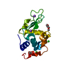 8cl5C 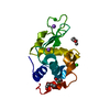 8cl6C  8cl7C  8cl8C  8clbC  8clcC 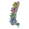 8cldC  8cleC  8clfC  8clgC  8clhC C: citing same article ( |
|---|---|
| Similar structure data |
-Links
 PDBj
PDBj


- Assembly
| Deposited unit | 
| ||||||||
|---|---|---|---|---|---|---|---|---|---|
| 1 |
| ||||||||
| Unit cell |
|
-Components
-Protein , 3 types, 3 molecules ABF
| #1: Protein | Mass: 48665.027 Da / Num. of mol.: 1 / Source method: isolated from a natural source / Source: (natural) |
|---|---|
| #2: Protein | Mass: 48391.410 Da / Num. of mol.: 1 / Source method: isolated from a natural source / Source: (natural) |
| #3: Protein | Mass: 16435.645 Da / Num. of mol.: 1 Source method: isolated from a genetically manipulated source Source: (gene. exp.) synthetic construct (others) / Production host:  |
-Non-polymers , 3 types, 148 molecules 


| #4: Chemical |  Mass: 523.180 Da / Num. of mol.: 2 / Source method: isolated from a natural source / Formula: C10H16N5O14P3 / Comment: GTP, energy-carrying molecule*YM Mass: 523.180 Da / Num. of mol.: 2 / Source method: isolated from a natural source / Formula: C10H16N5O14P3 / Comment: GTP, energy-carrying molecule*YM#5: Chemical | ChemComp-MG / |  Mass: 24.305 Da / Num. of mol.: 1 / Source method: obtained synthetically / Formula: Mg Mass: 24.305 Da / Num. of mol.: 1 / Source method: obtained synthetically / Formula: Mg#6: Water | ChemComp-HOH / | Mass: 18.015 Da / Num. of mol.: 145 / Source method: isolated from a natural source / Formula: H2O |
|---|
-Details
| Has ligand of interest | N |
|---|
-Experimental details
-Experiment
| Experiment | Method: X-RAY DIFFRACTION / Number of used crystals: 1 |
|---|
- Sample preparation
| Crystal | Density Matthews: 2.44 Å3/Da / Density % sol: 49.54 % |
|---|---|
| Crystal grow | Temperature: 293 K / Method: batch mode / pH: 5.5 Details: 21% PEG3000 (w/v), 0.2 M Ammonium Sulfate, 0.1 M Bis-Tris Methane |
-Data collection
| Diffraction | Mean temperature: 293 K / Serial crystal experiment: Y |
|---|---|
| Diffraction source | Source: FREE ELECTRON LASER / Site: SwissFEL ARAMIS / Beamline: ESA / Wavelength: 1.03 Å |
| Detector | Type: PSI JUNGFRAU 16M / Detector: PIXEL / Date: Feb 28, 2020 |
| Radiation | Protocol: SINGLE WAVELENGTH / Monochromatic (M) / Laue (L): M / Scattering type: x-ray |
| Radiation wavelength | Wavelength: 1.03 Å / Relative weight: 1 |
| Reflection | Resolution: 2.1→14.94 Å / Num. obs: 63162 / % possible obs: 100 % / Redundancy: 71.9 % / CC1/2: 0.931 / Net I/σ(I): 3.58 |
| Reflection shell | Resolution: 2.1→2.2 Å / Mean I/σ(I) obs: 0.52 / Num. unique obs: 6206 / CC1/2: 0.323 |
| Serial crystallography sample delivery | Method: injection |
| Serial crystallography sample delivery injection | Carrier solvent: Hydroxyethylcellulose / Description: High-viscosity injection / Injector diameter: 75 µm |
- Processing
| Software |
| |||||||||||||||||||||||||||||||||||||||||||||||||||||||||||||||||||||||||||||||||||||||||||
|---|---|---|---|---|---|---|---|---|---|---|---|---|---|---|---|---|---|---|---|---|---|---|---|---|---|---|---|---|---|---|---|---|---|---|---|---|---|---|---|---|---|---|---|---|---|---|---|---|---|---|---|---|---|---|---|---|---|---|---|---|---|---|---|---|---|---|---|---|---|---|---|---|---|---|---|---|---|---|---|---|---|---|---|---|---|---|---|---|---|---|---|---|
| Refinement | Method to determine structure: MOLECULAR REPLACEMENT / Resolution: 2.5→13 Å / SU ML: 0.3 / Cross valid method: FREE R-VALUE / σ(F): 1.34 / Phase error: 23.79 / Stereochemistry target values: ML
| |||||||||||||||||||||||||||||||||||||||||||||||||||||||||||||||||||||||||||||||||||||||||||
| Solvent computation | Shrinkage radii: 0.9 Å / VDW probe radii: 1.1 Å / Solvent model: FLAT BULK SOLVENT MODEL | |||||||||||||||||||||||||||||||||||||||||||||||||||||||||||||||||||||||||||||||||||||||||||
| Refinement step | Cycle: LAST / Resolution: 2.5→13 Å
| |||||||||||||||||||||||||||||||||||||||||||||||||||||||||||||||||||||||||||||||||||||||||||
| Refine LS restraints |
| |||||||||||||||||||||||||||||||||||||||||||||||||||||||||||||||||||||||||||||||||||||||||||
| LS refinement shell |
|
