 Movie
Movie Controller

[English] 日本語
 Yorodumi
- PDB-8cdq: Plasmodium falciparum Myosin A full-length, post-rigor state comp... -
+ Open data

- Basic information
| Entry | Database: PDB / ID: 8cdq | ||||||
|---|---|---|---|---|---|---|---|
| Title | Plasmodium falciparum Myosin A full-length, post-rigor state complexed to the inhibitor KNX-002 and Mg.ATP-gamma-S | ||||||
 Components Components |
| ||||||
 Keywords Keywords | MOTOR PROTEIN / Malaria / Plasmodium falciparum / Myosin A / KNX-002 / antimalarial treatment / molecular motors | ||||||
| Function / homology |  Function and homology information Function and homology informationpellicle / inner membrane pellicle complex / glideosome / myosin complex / myosin II complex / microfilament motor activity / cytoskeletal motor activity / actin filament organization / actin filament binding / actin cytoskeleton ...pellicle / inner membrane pellicle complex / glideosome / myosin complex / myosin II complex / microfilament motor activity / cytoskeletal motor activity / actin filament organization / actin filament binding / actin cytoskeleton / actin binding / calcium ion binding / ATP binding / membrane / plasma membrane / cytoplasm Similarity search - Function | ||||||
| Biological species |  | ||||||
| Method |  X-RAY DIFFRACTION / SYNCHROTRON / MOLECULAR REPLACEMENT / Resolution: 2.21 Å X-RAY DIFFRACTION / SYNCHROTRON / MOLECULAR REPLACEMENT / Resolution: 2.21 Å | ||||||
 Authors Authors | Moussaoui, D. / Robblee, J.P. / Robert-Paganin, J. / Auguin, D. / Fisher, F. / Fagnant, P.M. / MacFarlane, J.E. / Schaletzky, J. / Wehri, E. / Mueller-Dieckmann, C. ...Moussaoui, D. / Robblee, J.P. / Robert-Paganin, J. / Auguin, D. / Fisher, F. / Fagnant, P.M. / MacFarlane, J.E. / Schaletzky, J. / Wehri, E. / Mueller-Dieckmann, C. / Baum, J. / Trybus, K.M. / Houdusse, A. | ||||||
| Funding support |  United States, 1items United States, 1items
| ||||||
 Citation Citation | Journal: Nat Commun / Year: 2023 Title: Mechanism of small molecule inhibition of Plasmodium falciparum myosin A informs antimalarial drug design. Authors: Moussaoui, D. / Robblee, J.P. / Robert-Paganin, J. / Auguin, D. / Fisher, F. / Fagnant, P.M. / Macfarlane, J.E. / Schaletzky, J. / Wehri, E. / Mueller-Dieckmann, C. / Baum, J. / Trybus, K.M. / Houdusse, A. | ||||||
| History |
|
- Structure visualization
| Structure viewer | Molecule: MolmilJmol/JSmol |
|---|
- Downloads & links
-Download
| PDBx/mmCIF format | 8cdq.cif.gz | 463.4 KB | Display | PDBx/mmCIF format |
|---|---|---|---|---|
| PDB format | pdb8cdq.ent.gz | 370.5 KB | Display | PDB format |
| PDBx/mmJSON format | 8cdq.json.gz | Tree view | PDBx/mmJSON format | |
| Others |  Other downloads Other downloads |
-Validation report
| Arichive directory | https://data.pdbj.org/pub/pdb/validation_reports/cd/8cdqftp://data.pdbj.org/pub/pdb/validation_reports/cd/8cdq | HTTPS FTP |
|---|
-Related structure data
| Related structure data | 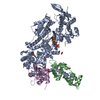 8a12C  8cdmC  6ycyS C: citing same article ( S: Starting model for refinement |
|---|---|
| Similar structure data |
-Links
 PDBj
PDBj


- Assembly
| Deposited unit | 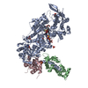
| ||||||||
|---|---|---|---|---|---|---|---|---|---|
| 1 |
| ||||||||
| Unit cell |
|
-Components
-Protein , 3 types, 3 molecules ABC
| #1: Protein | Mass: 92474.305 Da / Num. of mol.: 1 Source method: isolated from a genetically manipulated source Source: (gene. exp.) Strain: isolate 3D7 / Gene: MyoA, PF13_0233, PF3D7_1342600 / Production host:   Spodoptera frugiperda (fall armyworm) / References: UniProt: Q8IDR3 Spodoptera frugiperda (fall armyworm) / References: UniProt: Q8IDR3 |
|---|---|
| #2: Protein | Mass: 23510.738 Da / Num. of mol.: 1 Source method: isolated from a genetically manipulated source Source: (gene. exp.) Strain: isolate 3D7 / Gene: PF3D7_1246400 / Production host: Spodoptera frugiperda (fall armyworm) / References: UniProt: Q8I4W8 |
| #3: Protein | Mass: 15692.797 Da / Num. of mol.: 1 Source method: isolated from a genetically manipulated source Source: (gene. exp.) Strain: isolate 3D7 / Gene: PF3D7_1017500 / Production host: Spodoptera frugiperda (fall armyworm) / References: UniProt: Q8IJM4 |
-Non-polymers , 8 types, 317 molecules 


| #4: Chemical | ChemComp-GOL /  Mass: 92.094 Da / Num. of mol.: 4 / Source method: obtained synthetically / Formula: C3H8O3 Mass: 92.094 Da / Num. of mol.: 4 / Source method: obtained synthetically / Formula: C3H8O3#5: Chemical |  Mass: 96.063 Da / Num. of mol.: 2 / Source method: obtained synthetically / Formula: SO4 Mass: 96.063 Da / Num. of mol.: 2 / Source method: obtained synthetically / Formula: SO4#6: Chemical |  Mass: 62.068 Da / Num. of mol.: 2 / Source method: obtained synthetically / Formula: C2H6O2 Mass: 62.068 Da / Num. of mol.: 2 / Source method: obtained synthetically / Formula: C2H6O2#7: Chemical | ChemComp-PG4 / |  Mass: 194.226 Da / Num. of mol.: 1 / Source method: obtained synthetically / Formula: C8H18O5 / Comment: precipitant*YM Mass: 194.226 Da / Num. of mol.: 1 / Source method: obtained synthetically / Formula: C8H18O5 / Comment: precipitant*YM#8: Chemical | ChemComp-AGS / |  Mass: 523.247 Da / Num. of mol.: 1 / Source method: obtained synthetically / Formula: C10H16N5O12P3S / Feature type: SUBJECT OF INVESTIGATION / Comment: ATP-gamma-S, energy-carrying molecule analogue*YM Mass: 523.247 Da / Num. of mol.: 1 / Source method: obtained synthetically / Formula: C10H16N5O12P3S / Feature type: SUBJECT OF INVESTIGATION / Comment: ATP-gamma-S, energy-carrying molecule analogue*YM#9: Chemical | ChemComp-MG / |  Mass: 24.305 Da / Num. of mol.: 1 / Source method: obtained synthetically / Formula: Mg Mass: 24.305 Da / Num. of mol.: 1 / Source method: obtained synthetically / Formula: Mg#10: Chemical | ChemComp-KQ0 / |  Mass: 325.428 Da / Num. of mol.: 1 / Source method: obtained synthetically / Formula: C18H19N3OS / Feature type: SUBJECT OF INVESTIGATION Mass: 325.428 Da / Num. of mol.: 1 / Source method: obtained synthetically / Formula: C18H19N3OS / Feature type: SUBJECT OF INVESTIGATION#11: Water | ChemComp-HOH / | Mass: 18.015 Da / Num. of mol.: 305 / Source method: isolated from a natural source / Formula: H2O |
|---|
-Details
| Has ligand of interest | Y |
|---|---|
| Has protein modification | Y |
-Experimental details
-Experiment
| Experiment | Method: X-RAY DIFFRACTION / Number of used crystals: 1 |
|---|
- Sample preparation
| Crystal | Density Matthews: 3.36 Å3/Da / Density % sol: 63.35 % |
|---|---|
| Crystal grow | Temperature: 277.15 K / Method: vapor diffusion, hanging drop Details: 2.0 M ammonium sulfate, 0.1M sodium HEPES pH 7,5, 4% PEG400 |
-Data collection
| Diffraction | Mean temperature: 100 K / Serial crystal experiment: N |
|---|---|
| Diffraction source | Source: SYNCHROTRON / Site: ESRF  / Beamline: ID30B / Wavelength: 0.9687 Å / Beamline: ID30B / Wavelength: 0.9687 Å |
| Detector | Type: DECTRIS EIGER2 S 9M / Detector: PIXEL / Date: Dec 12, 2022 |
| Radiation | Protocol: SINGLE WAVELENGTH / Monochromatic (M) / Laue (L): M / Scattering type: x-ray |
| Radiation wavelength | Wavelength: 0.9687 Å / Relative weight: 1 |
| Reflection | Resolution: 2.21→95.08 Å / Num. obs: 57115 / % possible obs: 93.9 % / Redundancy: 7 % / CC1/2: 0.992 / Rmerge(I) obs: 0.199 / Net I/σ(I): 8.1 |
| Reflection shell | Resolution: 2.21→2.43 Å / Rmerge(I) obs: 1.476 / Mean I/σ(I) obs: 1.5 / Num. unique obs: 2857 / CC1/2: 0.546 / % possible all: 59.2 |
- Processing
| Software |
| |||||||||||||||||||||||||||||||||||||||||||||||||||||||||||||||||||||||||||||||||||||||||||||||||||||||||||||||||||||||||||||||||||||||||||||||||||
|---|---|---|---|---|---|---|---|---|---|---|---|---|---|---|---|---|---|---|---|---|---|---|---|---|---|---|---|---|---|---|---|---|---|---|---|---|---|---|---|---|---|---|---|---|---|---|---|---|---|---|---|---|---|---|---|---|---|---|---|---|---|---|---|---|---|---|---|---|---|---|---|---|---|---|---|---|---|---|---|---|---|---|---|---|---|---|---|---|---|---|---|---|---|---|---|---|---|---|---|---|---|---|---|---|---|---|---|---|---|---|---|---|---|---|---|---|---|---|---|---|---|---|---|---|---|---|---|---|---|---|---|---|---|---|---|---|---|---|---|---|---|---|---|---|---|---|---|---|
| Refinement | Method to determine structure: MOLECULAR REPLACEMENT Starting model: 6YCY Resolution: 2.21→95.08 Å / SU ML: 0.25 / Cross valid method: FREE R-VALUE / σ(F): 1.34 / Phase error: 30.38 / Stereochemistry target values: ML
| |||||||||||||||||||||||||||||||||||||||||||||||||||||||||||||||||||||||||||||||||||||||||||||||||||||||||||||||||||||||||||||||||||||||||||||||||||
| Solvent computation | Shrinkage radii: 0.9 Å / VDW probe radii: 1.11 Å / Solvent model: FLAT BULK SOLVENT MODEL | |||||||||||||||||||||||||||||||||||||||||||||||||||||||||||||||||||||||||||||||||||||||||||||||||||||||||||||||||||||||||||||||||||||||||||||||||||
| Refinement step | Cycle: LAST / Resolution: 2.21→95.08 Å
| |||||||||||||||||||||||||||||||||||||||||||||||||||||||||||||||||||||||||||||||||||||||||||||||||||||||||||||||||||||||||||||||||||||||||||||||||||
| Refine LS restraints |
| |||||||||||||||||||||||||||||||||||||||||||||||||||||||||||||||||||||||||||||||||||||||||||||||||||||||||||||||||||||||||||||||||||||||||||||||||||
| LS refinement shell |
| |||||||||||||||||||||||||||||||||||||||||||||||||||||||||||||||||||||||||||||||||||||||||||||||||||||||||||||||||||||||||||||||||||||||||||||||||||
| Refinement TLS params. | Method: refined / Origin x: 0.2041 Å / Origin y: -48.1242 Å / Origin z: 2.7952 Å
| |||||||||||||||||||||||||||||||||||||||||||||||||||||||||||||||||||||||||||||||||||||||||||||||||||||||||||||||||||||||||||||||||||||||||||||||||||
| Refinement TLS group | Selection details: all |
