 Movie
Movie Controller

[English] 日本語
 Yorodumi
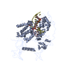
- PDB-8c59: CpG specific M.MpeI methyltransferase crystallized in the presenc... -
+ Open data

- Basic information
| Entry | Database: PDB / ID: 8c59 | |||||||||||||||||||||||||||
|---|---|---|---|---|---|---|---|---|---|---|---|---|---|---|---|---|---|---|---|---|---|---|---|---|---|---|---|---|
| Title | CpG specific M.MpeI methyltransferase crystallized in the presence of 5-bromocytosine (converted to 5mC) and 5-methylcytosine containing dsDNA | |||||||||||||||||||||||||||
 Components Components |
| |||||||||||||||||||||||||||
 Keywords Keywords | TRANSFERASE / M.MpeI / DNA methyltransferase / DNA / 5-methylcytosine / 5-bromocytosine / 5BrC / 5mC / CpG | |||||||||||||||||||||||||||
| Function / homology |  Function and homology information Function and homology informationDNA (cytosine-5-)-methyltransferase / DNA (cytosine-5-)-methyltransferase activity / DNA restriction-modification system / methylation Similarity search - Function | |||||||||||||||||||||||||||
| Biological species |  Malacoplasma penetrans HF-2 (bacteria) Malacoplasma penetrans HF-2 (bacteria)synthetic construct (others) | |||||||||||||||||||||||||||
| Method |  X-RAY DIFFRACTION / SYNCHROTRON / MOLECULAR REPLACEMENT / Resolution: 1.7 Å X-RAY DIFFRACTION / SYNCHROTRON / MOLECULAR REPLACEMENT / Resolution: 1.7 Å | |||||||||||||||||||||||||||
 Authors Authors | Wojciechowski, M. / Czapinska, H. / Krwawicz, J. / Rafalski, D. / Bochtler, M. | |||||||||||||||||||||||||||
| Funding support |  Poland, European Union, 8items Poland, European Union, 8items
| |||||||||||||||||||||||||||
 Citation Citation | Journal: Nucleic Acids Res. / Year: 2024 Title: Cytosine analogues as DNA methyltransferase substrates. Authors: Wojciechowski, M. / Czapinska, H. / Krwawicz, J. / Rafalski, D. / Bochtler, M. #1: Journal: Proc. Natl. Acad. Sci. U.S.A. / Year: 2013Title: CpG underrepresentation and the bacterial CpG-specific DNA methyltransferase M.MpeI. Authors: Wojciechowski, M. / Czapinska, H. / Bochtler, M. #2: Journal: Science / Year: 2012Title: Structure-based mechanistic insights into DNMT1-mediated maintenance DNA methylation. Authors: Song, J. / Teplova, M. / Ishibe-Murakami, S. / Patel, D.J. | |||||||||||||||||||||||||||
| History |
|
- Structure visualization
| Structure viewer | Molecule: MolmilJmol/JSmol |
|---|
- Downloads & links
-Download
| PDBx/mmCIF format | 8c59.cif.gz | 250.8 KB | Display | PDBx/mmCIF format |
|---|---|---|---|---|
| PDB format | pdb8c59.ent.gz | 198.6 KB | Display | PDB format |
| PDBx/mmJSON format | 8c59.json.gz | Tree view | PDBx/mmJSON format | |
| Others |  Other downloads Other downloads |
-Validation report
| Summary document | 8c59_validation.pdf.gz | 777.5 KB | Display | wwPDB validaton report |
|---|---|---|---|---|
| Full document | 8c59_full_validation.pdf.gz | 781.1 KB | Display | |
| Data in XML | 8c59_validation.xml.gz | 24.5 KB | Display | |
| Data in CIF | 8c59_validation.cif.gz | 37.8 KB | Display | |
| Arichive directory | https://data.pdbj.org/pub/pdb/validation_reports/c5/8c59ftp://data.pdbj.org/pub/pdb/validation_reports/c5/8c59 | HTTPS FTP |
-Related structure data
| Related structure data |  8c56C  8c57C  8c58C  4dkjS S: Starting model for refinement C: citing same article ( |
|---|---|
| Similar structure data |
-Links
 PDBj
PDBj


- Assembly
| Deposited unit | 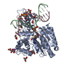
| ||||||||
|---|---|---|---|---|---|---|---|---|---|
| 1 |
| ||||||||
| Unit cell |
| ||||||||
| Components on special symmetry positions |
|
-Components
-Protein , 1 types, 1 molecules A
| #1: Protein | Mass: 46238.941 Da / Num. of mol.: 1 Source method: isolated from a genetically manipulated source Source: (gene. exp.) Malacoplasma penetrans HF-2 (bacteria) / Strain: HF-2 / Gene: MYPE4940 / Plasmid: pET-28a / Production host: |
|---|
-DNA chain , 2 types, 2 molecules BC
| #2: DNA chain | Mass: 4263.809 Da / Num. of mol.: 1 Source method: isolated from a genetically manipulated source Source: (gene. exp.) synthetic construct (others) / Production host: synthetic construct (others) |
|---|---|
| #3: DNA chain | Mass: 4325.829 Da / Num. of mol.: 1 Source method: isolated from a genetically manipulated source Source: (gene. exp.) synthetic construct (others) / Production host: synthetic construct (others) |
-Non-polymers , 6 types, 604 molecules 


| #4: Chemical | ChemComp-SAM /  Mass: 398.437 Da / Num. of mol.: 1 / Source method: obtained synthetically / Formula: C15H22N6O5S Mass: 398.437 Da / Num. of mol.: 1 / Source method: obtained synthetically / Formula: C15H22N6O5S | ||||||
|---|---|---|---|---|---|---|---|
| #5: Chemical | ChemComp-NA /  Mass: 22.990 Da / Num. of mol.: 1 / Source method: obtained synthetically / Formula: Na Mass: 22.990 Da / Num. of mol.: 1 / Source method: obtained synthetically / Formula: Na | ||||||
| #6: Chemical | ChemComp-GOL /  Mass: 92.094 Da / Num. of mol.: 9 / Source method: obtained synthetically / Formula: C3H8O3 Mass: 92.094 Da / Num. of mol.: 9 / Source method: obtained synthetically / Formula: C3H8O3#7: Chemical | ChemComp-CIT / |  Mass: 192.124 Da / Num. of mol.: 1 / Source method: obtained synthetically / Formula: C6H8O7 Mass: 192.124 Da / Num. of mol.: 1 / Source method: obtained synthetically / Formula: C6H8O7#8: Chemical | ChemComp-CO3 / |  Mass: 60.009 Da / Num. of mol.: 1 / Source method: obtained synthetically / Formula: CO3 Mass: 60.009 Da / Num. of mol.: 1 / Source method: obtained synthetically / Formula: CO3#9: Water | ChemComp-HOH / | Mass: 18.015 Da / Num. of mol.: 591 / Source method: isolated from a natural source / Formula: H2O |
-Details
| Has ligand of interest | Y |
|---|
-Experimental details
-Experiment
| Experiment | Method: X-RAY DIFFRACTION / Number of used crystals: 1 |
|---|
- Sample preparation
| Crystal | Density Matthews: 2.8 Å3/Da / Density % sol: 56.11 % |
|---|---|
| Crystal grow | Temperature: 294 K / Method: vapor diffusion, sitting drop / pH: 5.6 Details: 10% PEG 3350, 150 mM NaCl, 50 mM sodium citrate (final pH, 5.6). For cryoprotection glycerol was added to 25% v/v |
-Data collection
| Diffraction | Mean temperature: 100 K / Serial crystal experiment: N |
|---|---|
| Diffraction source | Source: SYNCHROTRON / Site: PETRA III, EMBL c/o DESY  / Beamline: P13 (MX1) / Wavelength: 0.9116 Å / Beamline: P13 (MX1) / Wavelength: 0.9116 Å |
| Detector | Type: DECTRIS PILATUS 6M-F / Detector: PIXEL / Date: Sep 5, 2016 |
| Radiation | Protocol: SINGLE WAVELENGTH / Monochromatic (M) / Laue (L): M / Scattering type: x-ray |
| Radiation wavelength | Wavelength: 0.9116 Å / Relative weight: 1 |
| Reflection | Resolution: 1.7→20 Å / Num. obs: 69838 / % possible obs: 99.8 % / Redundancy: 8.78 % / Biso Wilson estimate: 38.44 Å2 / CC1/2: 1 / Rrim(I) all: 0.041 / Rsym value: 0.039 / Net I/σ(I): 27.87 |
| Reflection shell | Resolution: 1.7→1.8 Å / Redundancy: 8.52 % / Mean I/σ(I) obs: 2.15 / Num. unique obs: 11063 / CC1/2: 0.822 / Rrim(I) all: 0.98 / Rsym value: 0.919 / % possible all: 99.8 |
- Processing
| Software |
| ||||||||||||||||||||||||||||||||||||||||||||||||||||||||||||||||||||||||||||||||||||||||||||||||||||||||||||||||||||||||||||||||||||||||||||||||||||||||||||||||||||||||||||||||||||||
|---|---|---|---|---|---|---|---|---|---|---|---|---|---|---|---|---|---|---|---|---|---|---|---|---|---|---|---|---|---|---|---|---|---|---|---|---|---|---|---|---|---|---|---|---|---|---|---|---|---|---|---|---|---|---|---|---|---|---|---|---|---|---|---|---|---|---|---|---|---|---|---|---|---|---|---|---|---|---|---|---|---|---|---|---|---|---|---|---|---|---|---|---|---|---|---|---|---|---|---|---|---|---|---|---|---|---|---|---|---|---|---|---|---|---|---|---|---|---|---|---|---|---|---|---|---|---|---|---|---|---|---|---|---|---|---|---|---|---|---|---|---|---|---|---|---|---|---|---|---|---|---|---|---|---|---|---|---|---|---|---|---|---|---|---|---|---|---|---|---|---|---|---|---|---|---|---|---|---|---|---|---|---|---|
| Refinement | Method to determine structure: MOLECULAR REPLACEMENT Starting model: 4dkj Resolution: 1.7→19.95 Å / Cor.coef. Fo:Fc: 0.973 / Cor.coef. Fo:Fc free: 0.966 / SU B: 4.723 / SU ML: 0.068 / Cross valid method: THROUGHOUT / ESU R: 0.091 / ESU R Free: 0.089 / Stereochemistry target values: MAXIMUM LIKELIHOOD Details: HYDROGEN ATOMS HAVE BEEN ADDED IN THE RIDING POSITIONS. U VALUES : WITH TLS ADDED. IONS AND SOLVENT MOLECULES HAVE BEEN MODELLED TENTATIVELY. S-ADENOSYLMETHIONINE (SAM) HAS BEEN MODELLED IN ...Details: HYDROGEN ATOMS HAVE BEEN ADDED IN THE RIDING POSITIONS. U VALUES : WITH TLS ADDED. IONS AND SOLVENT MOLECULES HAVE BEEN MODELLED TENTATIVELY. S-ADENOSYLMETHIONINE (SAM) HAS BEEN MODELLED IN THE CO-SUBSTRATE BINDING POCKET BUT ONLY PARTS OF THE MOLECULE CORRESPONDING TO ITS BASE AND AMINO ACID END ARE RESOLVED IN THE ELECTRON DENSITY. THE CENTRAL PART OF THE CO-SUBSTRATE COULD NOT BE UNAMBIGUOUSLY TRACED, WHICH MAY BE DUE TO ITS DECOMPOSITION TO HOMOSERINE LACTONE (HSL) AND METHYLTHIOADENOSINE (MTA). 5-BROMOCYTOSINE CONTAINING DNA HAS BEEN USED FOR CRYSTALLIZATION BUT 5-METHYLCYTOSINE IS OBSERVED IN THE CRYSTALS IN THE M.MPEI SUBSTRATE BINDING POCKET WHICH INDICATES THAT THE REACTION MUST HAVE TAKEN PLACE EITHER IN SOLUTION PRIOR TO CRYSTALLIZATION OR IN THE CRYSTAL. THE RESIDUAL DIFFERENCE DENSITY NEXT TO THE SUBSTRATE BASE LIKELY RESULTS FROM THE RESIDUAL PRESENCE OF THE ACTIVE SITE LOOP IN THE "IN" CONFORMATION BUT THE OCCUPANCY OF THIS CONFORMER WAS TOO LOW TO BE INCLUDED IN THE MODEL. Since the pH of the crystallization drop (reservoir buffer pH 5.6) was close to the pK for the HCO3-/CO32-+H+ equilibrium (pKa=6.1), a mixture of carbonate and hydrogencarbonate is likely present in the crystal.
| ||||||||||||||||||||||||||||||||||||||||||||||||||||||||||||||||||||||||||||||||||||||||||||||||||||||||||||||||||||||||||||||||||||||||||||||||||||||||||||||||||||||||||||||||||||||
| Solvent computation | Ion probe radii: 0.8 Å / Shrinkage radii: 0.8 Å / VDW probe radii: 1.2 Å / Solvent model: MASK | ||||||||||||||||||||||||||||||||||||||||||||||||||||||||||||||||||||||||||||||||||||||||||||||||||||||||||||||||||||||||||||||||||||||||||||||||||||||||||||||||||||||||||||||||||||||
| Displacement parameters | Biso mean: 38.536 Å2
| ||||||||||||||||||||||||||||||||||||||||||||||||||||||||||||||||||||||||||||||||||||||||||||||||||||||||||||||||||||||||||||||||||||||||||||||||||||||||||||||||||||||||||||||||||||||
| Refinement step | Cycle: 1 / Resolution: 1.7→19.95 Å
| ||||||||||||||||||||||||||||||||||||||||||||||||||||||||||||||||||||||||||||||||||||||||||||||||||||||||||||||||||||||||||||||||||||||||||||||||||||||||||||||||||||||||||||||||||||||
| Refine LS restraints |
|