 Movie
Movie Controller

[English] 日本語
 Yorodumi
- PDB-8c16: Crystal structure of asymmetric ferredoxin/flavodoxin NADP+ oxido... -
+ Open data

- Basic information
| Entry | Database: PDB / ID: 8c16 | |||||||||
|---|---|---|---|---|---|---|---|---|---|---|
| Title | Crystal structure of asymmetric ferredoxin/flavodoxin NADP+ oxidoreductase 2 (FNR2) H326V mutant from Bacillus cereus | |||||||||
 Components Components | Ferredoxin--NADP reductase | |||||||||
 Keywords Keywords | OXIDOREDUCTASE / FERREDOXIN/FLAVODOXIN REDUCTASE / ELECTRON TRANSFER / FAD / FLAVOPROTEIN / MUTANT | |||||||||
| Function / homology |  Function and homology information Function and homology informationferredoxin-NADP+ reductase / ferredoxin-NADP+ reductase activity / thioredoxin-disulfide reductase (NADPH) activity / cell redox homeostasis / NADP binding / flavin adenine dinucleotide binding Similarity search - Function | |||||||||
| Biological species |  | |||||||||
| Method |  X-RAY DIFFRACTION / SYNCHROTRON / MOLECULAR REPLACEMENT / Resolution: 4.2 Å X-RAY DIFFRACTION / SYNCHROTRON / MOLECULAR REPLACEMENT / Resolution: 4.2 Å | |||||||||
 Authors Authors | Rugtveit, A.K. / Hammerstad, M. / Hersleth, H.-P. | |||||||||
| Funding support |  Norway, 2items Norway, 2items
| |||||||||
 Citation Citation | Journal: Antioxidants / Year: 2023 Title: Functional Diversity of Homologous Oxidoreductases-Tuning of Substrate Specificity by a FAD-Stacking Residue for Iron Acquisition and Flavodoxin Reduction. Authors: Hammerstad, M. / Rugtveit, A.K. / Dahlen, S. / Andersen, H.K. / Hersleth, H.P. | |||||||||
| History |
|
- Structure visualization
| Structure viewer | Molecule: MolmilJmol/JSmol |
|---|
- Downloads & links
-Download
| PDBx/mmCIF format | 8c16.cif.gz | 318.1 KB | Display | PDBx/mmCIF format |
|---|---|---|---|---|
| PDB format | pdb8c16.ent.gz | 207.2 KB | Display | PDB format |
| PDBx/mmJSON format | 8c16.json.gz | Tree view | PDBx/mmJSON format | |
| Others |  Other downloads Other downloads |
-Validation report
| Arichive directory | https://data.pdbj.org/pub/pdb/validation_reports/c1/8c16ftp://data.pdbj.org/pub/pdb/validation_reports/c1/8c16 | HTTPS FTP |
|---|
-Related structure data
| Related structure data |  8avhC 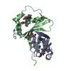 8aviC  8c3mC  6gasS S: Starting model for refinement C: citing same article ( |
|---|---|
| Similar structure data |
-Links
 PDBj
PDBj


- Assembly
| Deposited unit | 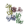
| ||||||||||||||||||||||||||||||||||||||||||||||
|---|---|---|---|---|---|---|---|---|---|---|---|---|---|---|---|---|---|---|---|---|---|---|---|---|---|---|---|---|---|---|---|---|---|---|---|---|---|---|---|---|---|---|---|---|---|---|---|
| 1 | 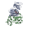
| ||||||||||||||||||||||||||||||||||||||||||||||
| 2 | 
| ||||||||||||||||||||||||||||||||||||||||||||||
| Unit cell |
| ||||||||||||||||||||||||||||||||||||||||||||||
| Noncrystallographic symmetry (NCS) | NCS domain:
NCS domain segments: Component-ID: 1 / Ens-ID: ens_1 / Beg auth comp-ID: ASN / Beg label comp-ID: ASN / End auth comp-ID: PRO / End label comp-ID: PRO / Auth seq-ID: 6 - 318 / Label seq-ID: 6 - 318
NCS oper:
|
-Components
| #1: Protein | Mass: 36754.094 Da / Num. of mol.: 4 / Mutation: H326V Source method: isolated from a genetically manipulated source Source: (gene. exp.) #2: Chemical |   Mass: 785.550 Da / Num. of mol.: 2 / Source method: obtained synthetically / Formula: C27H33N9O15P2 / Feature type: SUBJECT OF INVESTIGATION / Comment: FAD*YM Mass: 785.550 Da / Num. of mol.: 2 / Source method: obtained synthetically / Formula: C27H33N9O15P2 / Feature type: SUBJECT OF INVESTIGATION / Comment: FAD*YMHas ligand of interest | Y | |
|---|
-Experimental details
-Experiment
| Experiment | Method: X-RAY DIFFRACTION / Number of used crystals: 1 |
|---|
- Sample preparation
| Crystal | Density Matthews: 2.8 Å3/Da / Density % sol: 56.1 % |
|---|---|
| Crystal grow | Temperature: 298 K / Method: vapor diffusion, sitting drop / pH: 8.5 Details: 24 mg/mL protein (1:1) 10% w/v PEG 4000, 20% v/v glycerol, 0.02 M sodium l-glutamate, 0.02 M dl-alanine, 0.02 M glycine, 0.02 M dl-lysine HCl, 0.02 M dl-serine, 0.1 M bicine/Trizma base pH 8.5 |
-Data collection
| Diffraction | Mean temperature: 100 K / Serial crystal experiment: N |
|---|---|
| Diffraction source | Source: SYNCHROTRON / Site: ESRF  / Beamline: ID30B / Wavelength: 0.976254 Å / Beamline: ID30B / Wavelength: 0.976254 Å |
| Detector | Type: DECTRIS PILATUS3 6M / Detector: PIXEL / Date: Apr 18, 2021 |
| Radiation | Protocol: SINGLE WAVELENGTH / Monochromatic (M) / Laue (L): M / Scattering type: x-ray |
| Radiation wavelength | Wavelength: 0.976254 Å / Relative weight: 1 |
| Reflection | Resolution: 4.2→75.48 Å / Num. obs: 11894 / % possible obs: 95.8 % / Redundancy: 4 % / Biso Wilson estimate: 157.81 Å2 / CC1/2: 0.994 / Rmerge(I) obs: 0.177 / Rpim(I) all: 0.093 / Rrim(I) all: 0.202 / Χ2: 1.01 / Net I/σ(I): 5.6 |
| Reflection shell | Resolution: 4.2→4.7 Å / Redundancy: 4.2 % / Rmerge(I) obs: 0.962 / Mean I/σ(I) obs: 1.7 / Num. unique obs: 3352 / CC1/2: 0.552 / Rpim(I) all: 0.499 / Rrim(I) all: 1.092 / Χ2: 1.02 / % possible all: 96.8 |
- Processing
| Software |
| |||||||||||||||||||||||||||||||||||
|---|---|---|---|---|---|---|---|---|---|---|---|---|---|---|---|---|---|---|---|---|---|---|---|---|---|---|---|---|---|---|---|---|---|---|---|---|
| Refinement | Method to determine structure: MOLECULAR REPLACEMENT Starting model: 6GAS Resolution: 4.2→75.48 Å / SU ML: 0.6695 / Cross valid method: FREE R-VALUE / σ(F): 1.34 / Phase error: 37.2221 Stereochemistry target values: GeoStd + Monomer Library + CDL v1.2
| |||||||||||||||||||||||||||||||||||
| Solvent computation | Shrinkage radii: 0.9 Å / VDW probe radii: 1.1 Å / Solvent model: FLAT BULK SOLVENT MODEL | |||||||||||||||||||||||||||||||||||
| Displacement parameters | Biso mean: 185.98 Å2 | |||||||||||||||||||||||||||||||||||
| Refinement step | Cycle: LAST / Resolution: 4.2→75.48 Å
| |||||||||||||||||||||||||||||||||||
| Refine LS restraints |
| |||||||||||||||||||||||||||||||||||
| Refine LS restraints NCS |
| |||||||||||||||||||||||||||||||||||
| LS refinement shell |
|
