 Movie
Movie Controller

[English] 日本語
 Yorodumi
- PDB-8bw0: Structure of CEACAM5 A3-B3 domain in Complex with Tusamitamab Fab -
+ Open data

- Basic information
| Entry | Database: PDB / ID: 8bw0 | ||||||
|---|---|---|---|---|---|---|---|
| Title | Structure of CEACAM5 A3-B3 domain in Complex with Tusamitamab Fab | ||||||
 Components Components |
| ||||||
 Keywords Keywords | CELL ADHESION / CEACAM5 / Tusamitamab / Cancer / cryo-EM / small molecular weight / Fab / A3-B3 / human membrane protein | ||||||
| Function / homology |  Function and homology information Function and homology informationGPI anchor binding / homotypic cell-cell adhesion / negative regulation of myotube differentiation / Post-translational modification: synthesis of GPI-anchored proteins / heterophilic cell-cell adhesion / homophilic cell-cell adhesion / negative regulation of anoikis / side of membrane / Cell surface interactions at the vascular wall / basolateral plasma membrane ...GPI anchor binding / homotypic cell-cell adhesion / negative regulation of myotube differentiation / Post-translational modification: synthesis of GPI-anchored proteins / heterophilic cell-cell adhesion / homophilic cell-cell adhesion / negative regulation of anoikis / side of membrane / Cell surface interactions at the vascular wall / basolateral plasma membrane / apical plasma membrane / apoptotic process / negative regulation of apoptotic process / cell surface / protein homodimerization activity / extracellular exosome / extracellular region / membrane / identical protein binding / plasma membrane Similarity search - Function | ||||||
| Biological species |   Homo sapiens (human) Homo sapiens (human) | ||||||
| Method | ELECTRON MICROSCOPY / single particle reconstruction / cryo EM / Resolution: 3.11 Å | ||||||
 Authors Authors | Kumar, A. / Bertrand, T. / Rapisarda, C. / Rak, A. | ||||||
| Funding support |  France, 1items France, 1items
| ||||||
 Citation Citation | Journal: Nat Commun / Year: 2024 Title: Structural insights into epitope-paratope interactions of a monoclonal antibody targeting CEACAM5-expressing tumors. Authors: Anand Kumar / Francis Duffieux / Marie Gagnaire / Chiara Rapisarda / Thomas Bertrand / Alexey Rak / Abstract: Carcinoembryonic antigen-related cell adhesion molecules (CEACAMs) are overexpressed in some tumor types. The antibody-drug conjugate tusamitamab ravtansine specifically recognizes the A3-B3 domains ...Carcinoembryonic antigen-related cell adhesion molecules (CEACAMs) are overexpressed in some tumor types. The antibody-drug conjugate tusamitamab ravtansine specifically recognizes the A3-B3 domains of human CEACAM5 (hCEACAM5). To understand this specificity, here we map the epitope-paratope interface between the A3-B3 domains of hCEACAM5 (hCEACAM5) and the antigen-binding fragment of tusamitamab (tusa Fab). We use hydrogen/deuterium exchange mass spectrometry to identify the tusa Fab paratope, which involves heavy chain (HC) residues 101-109 and light chain residues 48-54 and 88-104. Using surface plasmon resonance, we demonstrate that alanine variants of HC residues 96-108 abolish binding to hCEACAM5, suggesting that these residues are critical for tusa-Fab-antigen complex formation. The cryogenic electron microscopy structure of the hCEACAM5- tusa Fab complex (3.11 Å overall resolution) reveals a discontinuous epitope involving residues in the A3-B3 domains and an N-linked mannose at residue Asn612. Conformational constraints on the epitope-paratope interface enable tusamitamab to target hCEACAM5 and distinguish CEACAM5 from other CEACAMs. #1: Journal: Res Sq / Year: 2023Title: Structural insights into epitope-paratope interactions of monoclonal antibody targeting CEACAM5-expressing tumors Authors: Rak, A. / Kumar, A. / Duffi, F. / Gagnaire, M. / Rapisarda, C. / Bertrand, T. | ||||||
| History |
|
- Structure visualization
| Structure viewer | Molecule: MolmilJmol/JSmol |
|---|
- Downloads & links
-Download
| PDBx/mmCIF format | 8bw0.cif.gz | 91.7 KB | Display | PDBx/mmCIF format |
|---|---|---|---|---|
| PDB format | pdb8bw0.ent.gz | 64 KB | Display | PDB format |
| PDBx/mmJSON format | 8bw0.json.gz | Tree view | PDBx/mmJSON format | |
| Others |  Other downloads Other downloads |
-Validation report
| Arichive directory | https://data.pdbj.org/pub/pdb/validation_reports/bw/8bw0ftp://data.pdbj.org/pub/pdb/validation_reports/bw/8bw0 | HTTPS FTP |
|---|
-Related structure data
| Related structure data |  16279MC M: map data used to model this data C: citing same article ( |
|---|---|
| Similar structure data |
-Links
 PDBj
PDBj


- Assembly
| Deposited unit | 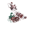
|
|---|---|
| 1 |
|
-Components
-Protein , 1 types, 1 molecules C
| #3: Protein | Mass: 20883.041 Da / Num. of mol.: 1 Source method: isolated from a genetically manipulated source Details: A3-B3 domain / Source: (gene. exp.) Homo sapiens (human) / Gene: CEACAM5, CEA / Cell line (production host): HEK293FS / Production host: Homo sapiens (human) / References: UniProt: P06731 |
|---|
-Antibody , 2 types, 2 molecules HL
| #1: Antibody | Mass: 24833.652 Da / Num. of mol.: 1 Source method: isolated from a genetically manipulated source Source: (gene. exp.) Homo sapiens (human) |
|---|---|
| #2: Antibody | Mass: 23497.072 Da / Num. of mol.: 1 Source method: isolated from a genetically manipulated source Source: (gene. exp.) Homo sapiens (human) |
-Sugars , 3 types, 6 molecules 
| #4: Polysaccharide | Source method: isolated from a genetically manipulated source #5: Polysaccharide | alpha-D-mannopyranose-(1-3)-[alpha-D-mannopyranose-(1-6)]beta-D-mannopyranose-(1-4)-2-acetamido-2- ...alpha-D-mannopyranose-(1-3)-[alpha-D-mannopyranose-(1-6)]beta-D-mannopyranose-(1-4)-2-acetamido-2-deoxy-beta-D-glucopyranose-(1-4)-[alpha-L-fucopyranose-(1-6)]2-acetamido-2-deoxy-beta-D-glucopyranose | Source method: isolated from a genetically manipulated source #6: Sugar |  Type: D-saccharide, beta linking / Mass: 221.208 Da / Num. of mol.: 3 / Source method: obtained synthetically / Formula: C8H15NO6 Type: D-saccharide, beta linking / Mass: 221.208 Da / Num. of mol.: 3 / Source method: obtained synthetically / Formula: C8H15NO6 |
|---|
-Details
| Has ligand of interest | N |
|---|---|
| Has protein modification | Y |
-Experimental details
-Experiment
| Experiment | Method: ELECTRON MICROSCOPY |
|---|---|
| EM experiment | Aggregation state: PARTICLE / 3D reconstruction method: single particle reconstruction |
- Sample preparation
| Component | Name: hCEACAM5-A3B3-Tusamitamab Fab complex / Type: COMPLEX Details: Human CEACAM5 a3-B3 domain in the comlex with the Tusamitamab Fab Entity ID: #1-#3 / Source: RECOMBINANT |
|---|---|
| Molecular weight | Value: 0.07 MDa / Experimental value: NO |
| Source (natural) | Organism: Homo sapiens (human) |
| Source (recombinant) | Organism: Homo sapiens (human) / Cell: HEK293FS |
| Buffer solution | pH: 7.4 / Details: Dulbeccos phosphate buffered saline |
| Buffer component | Conc.: 1 X / Name: D-PBS |
| Specimen | Conc.: 0.87 mg/ml / Embedding applied: NO / Shadowing applied: NO / Staining applied: NO / Vitrification applied: YES |
| Specimen support | Grid material: GOLD / Grid mesh size: 300 divisions/in. / Grid type: UltrAuFoil R0./1 |
| Vitrification | Instrument: FEI VITROBOT MARK IV / Cryogen name: ETHANE / Humidity: 100 % / Chamber temperature: 277.15 K |
- Electron microscopy imaging
| Microscopy | Model: TFS GLACIOS |
|---|---|
| Electron gun | Electron source:  FIELD EMISSION GUN / Accelerating voltage: 200 kV / Illumination mode: OTHER FIELD EMISSION GUN / Accelerating voltage: 200 kV / Illumination mode: OTHER |
| Electron lens | Mode: DARK FIELD / Nominal magnification: 240000 X / Nominal defocus max: 2200 nm / Nominal defocus min: 600 nm / Cs: 2.7 mm / C2 aperture diameter: 30 µm / Alignment procedure: COMA FREE |
| Specimen holder | Cryogen: NITROGEN |
| Image recording | Average exposure time: 4.72 sec. / Electron dose: 62 e/Å2 / Film or detector model: FEI FALCON IV (4k x 4k) / Num. of grids imaged: 1 / Num. of real images: 5072 |
- Processing
| Software | Name: PHENIX / Version: 1.19.2_4158: / Classification: refinement | ||||||||||||||||||||||||||||||||||||
|---|---|---|---|---|---|---|---|---|---|---|---|---|---|---|---|---|---|---|---|---|---|---|---|---|---|---|---|---|---|---|---|---|---|---|---|---|---|
| EM software |
| ||||||||||||||||||||||||||||||||||||
| CTF correction | Type: PHASE FLIPPING ONLY | ||||||||||||||||||||||||||||||||||||
| Particle selection | Num. of particles selected: 2614488 / Details: Picked using blob picker | ||||||||||||||||||||||||||||||||||||
| 3D reconstruction | Resolution: 3.11 Å / Resolution method: FSC 0.143 CUT-OFF / Num. of particles: 213470 / Num. of class averages: 1 / Symmetry type: POINT | ||||||||||||||||||||||||||||||||||||
| Atomic model building | Protocol: AB INITIO MODEL | ||||||||||||||||||||||||||||||||||||
| Refine LS restraints |
|
