 Movie
Movie Controller

[English] 日本語
 Yorodumi
- EMDB-16279: Structure of CEACAM5 A3-B3 domain in Complex with Tusamitamab Fab -
+ Open data

- Basic information
| Entry |  | |||||||||
|---|---|---|---|---|---|---|---|---|---|---|
| Title | Structure of CEACAM5 A3-B3 domain in Complex with Tusamitamab Fab | |||||||||
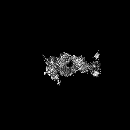
 Map data Map data | Cryo EM map for the complex | |||||||||
 Sample Sample |
| |||||||||
 Keywords Keywords | CEACAM5 / Tusamitamab / Cancer / cell adhesion / cryo-EM / small molecular weight / Fab / A3-B3 / human membrane protein | |||||||||
| Function / homology |  Function and homology information Function and homology informationGPI anchor binding / homotypic cell-cell adhesion / negative regulation of myotube differentiation / Post-translational modification: synthesis of GPI-anchored proteins / heterophilic cell-cell adhesion / homophilic cell-cell adhesion / negative regulation of anoikis / side of membrane / Cell surface interactions at the vascular wall / basolateral plasma membrane ...GPI anchor binding / homotypic cell-cell adhesion / negative regulation of myotube differentiation / Post-translational modification: synthesis of GPI-anchored proteins / heterophilic cell-cell adhesion / homophilic cell-cell adhesion / negative regulation of anoikis / side of membrane / Cell surface interactions at the vascular wall / basolateral plasma membrane / apical plasma membrane / apoptotic process / negative regulation of apoptotic process / cell surface / protein homodimerization activity / extracellular exosome / extracellular region / membrane / identical protein binding / plasma membrane Similarity search - Function | |||||||||
| Biological species |  Homo sapiens (human) / Homo sapiens (human) /  | |||||||||
| Method | single particle reconstruction / cryo EM / Resolution: 3.11 Å | |||||||||
 Authors Authors | Kumar A / Bertrand T / Rapisarda C / Rak A | |||||||||
| Funding support |  France, 1 items France, 1 items
| |||||||||
 Citation Citation | Journal: Nat Commun / Year: 2024 Title: Structural insights into epitope-paratope interactions of a monoclonal antibody targeting CEACAM5-expressing tumors. Authors: Anand Kumar / Francis Duffieux / Marie Gagnaire / Chiara Rapisarda / Thomas Bertrand / Alexey Rak / Abstract: Carcinoembryonic antigen-related cell adhesion molecules (CEACAMs) are overexpressed in some tumor types. The antibody-drug conjugate tusamitamab ravtansine specifically recognizes the A3-B3 domains ...Carcinoembryonic antigen-related cell adhesion molecules (CEACAMs) are overexpressed in some tumor types. The antibody-drug conjugate tusamitamab ravtansine specifically recognizes the A3-B3 domains of human CEACAM5 (hCEACAM5). To understand this specificity, here we map the epitope-paratope interface between the A3-B3 domains of hCEACAM5 (hCEACAM5) and the antigen-binding fragment of tusamitamab (tusa Fab). We use hydrogen/deuterium exchange mass spectrometry to identify the tusa Fab paratope, which involves heavy chain (HC) residues 101-109 and light chain residues 48-54 and 88-104. Using surface plasmon resonance, we demonstrate that alanine variants of HC residues 96-108 abolish binding to hCEACAM5, suggesting that these residues are critical for tusa-Fab-antigen complex formation. The cryogenic electron microscopy structure of the hCEACAM5- tusa Fab complex (3.11 Å overall resolution) reveals a discontinuous epitope involving residues in the A3-B3 domains and an N-linked mannose at residue Asn612. Conformational constraints on the epitope-paratope interface enable tusamitamab to target hCEACAM5 and distinguish CEACAM5 from other CEACAMs. #1: Journal: Res Sq / Year: 2023Title: Structural insights into epitope-paratope interactions of monoclonal antibody targeting CEACAM5-expressing tumors Authors: Rak A / Kumar A / Duffi F / Gagnaire M / Rapisarda C / Bertrand T | |||||||||
| History |
|
- Structure visualization
| Supplemental images |
|---|

- Downloads & links
-EMDB archive
| Map data | emd_16279.map.gz | 58.3 MB | EMDB map data format | |
|---|---|---|---|---|
| Header (meta data) | emd-16279-v30.xmlemd-16279.xml | 20.2 KB 20.2 KB | Display Display | EMDB header |
| Images |  emd_16279.png emd_16279.png | 80.5 KB | ||
| Filedesc metadata | emd-16279.cif.gz | 6.5 KB | ||
| Others | emd_16279_half_map_1.map.gzemd_16279_half_map_2.map.gz | 59.5 MB 59.5 MB | ||
| Archive directory |  http://ftp.pdbj.org/pub/emdb/structures/EMD-16279ftp://ftp.pdbj.org/pub/emdb/structures/EMD-16279 http://ftp.pdbj.org/pub/emdb/structures/EMD-16279ftp://ftp.pdbj.org/pub/emdb/structures/EMD-16279 | HTTPS FTP |
-Related structure data
| Related structure data |  8bw0MC M: atomic model generated by this map C: citing same article ( |
|---|---|
| Similar structure data |
-Links
| EMDB pages | EMDB (EBI/PDBe) / EMDataResource |
|---|---|
| Related items in Molecule of the Month |


-Map
| File | Download / File: emd_16279.map.gz / Format: CCP4 / Size: 64 MB / Type: IMAGE STORED AS FLOATING POINT NUMBER (4 BYTES) | ||||||||||||||||||||||||||||||||||||
|---|---|---|---|---|---|---|---|---|---|---|---|---|---|---|---|---|---|---|---|---|---|---|---|---|---|---|---|---|---|---|---|---|---|---|---|---|---|
| Annotation | Cryo EM map for the complex | ||||||||||||||||||||||||||||||||||||
| Projections & slices | Image control
Images are generated by Spider. | ||||||||||||||||||||||||||||||||||||
| Voxel size | X=Y=Z: 1.16 Å | ||||||||||||||||||||||||||||||||||||
| Density |
| ||||||||||||||||||||||||||||||||||||
| Symmetry | Space group: 1 | ||||||||||||||||||||||||||||||||||||
| Details | EMDB XML:
|
 X (Sec.)
 Y (Row.)
 Z (Col.)


-Supplemental data
-Half map: Half map A
| File | emd_16279_half_map_1.map | ||||||||||||
|---|---|---|---|---|---|---|---|---|---|---|---|---|---|
| Annotation | Half map A | ||||||||||||
| Projections & Slices |
| ||||||||||||
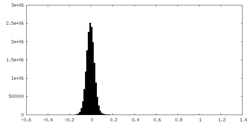
| Density Histograms |


-Half map: Half map B
| File | emd_16279_half_map_2.map | ||||||||||||
|---|---|---|---|---|---|---|---|---|---|---|---|---|---|
| Annotation | Half map B | ||||||||||||
| Projections & Slices |
| ||||||||||||
| Density Histograms |


- Sample components
-Entire : hCEACAM5-A3B3-Tusamitamab Fab complex
| Entire | Name: hCEACAM5-A3B3-Tusamitamab Fab complex |
|---|---|
| Components |
|
-Supramolecule #1: hCEACAM5-A3B3-Tusamitamab Fab complex
| Supramolecule | Name: hCEACAM5-A3B3-Tusamitamab Fab complex / type: complex / ID: 1 / Parent: 0 / Macromolecule list: #1-#3 Details: Human CEACAM5 a3-B3 domain in the comlex with the Tusamitamab Fab |
|---|---|
| Source (natural) | Organism: Homo sapiens (human) |
| Molecular weight | Theoretical: 70 KDa |
-Macromolecule #1: Tusamitamab Fab heavy Chain
| Macromolecule | Name: Tusamitamab Fab heavy Chain / type: protein_or_peptide / ID: 1 / Number of copies: 1 / Enantiomer: LEVO |
|---|---|
| Source (natural) | Organism: |
| Molecular weight | Theoretical: 24.833652 KDa |
| Recombinant expression | Organism: Homo sapiens (human) |
| Sequence | String: EVQLQESGPG LVKPGGSLSL SCAASGFVFS SYDMSWVRQT PERGLEWVAY ISSGGGITYA PSTVKGRFTV SRDNAKNTLY LQMNSLTSE DTAVYYCAAH YFGSSGPFAY WGQGTLVTVS SASTKGPSVF PLAPSSKSTS GGTAALGCLV KDYFPEPVTV S WNSGALTS ...String: EVQLQESGPG LVKPGGSLSL SCAASGFVFS SYDMSWVRQT PERGLEWVAY ISSGGGITYA PSTVKGRFTV SRDNAKNTLY LQMNSLTSE DTAVYYCAAH YFGSSGPFAY WGQGTLVTVS SASTKGPSVF PLAPSSKSTS GGTAALGCLV KDYFPEPVTV S WNSGALTS GVHTFPAVLQ SSGLYSLSSV VTVPSSSLGT QTYICNVNHK PSNTKVDKKV EPKSCDKTHT HHHHHH |
-Macromolecule #2: Tusamitamab Light Chain
| Macromolecule | Name: Tusamitamab Light Chain / type: protein_or_peptide / ID: 2 / Number of copies: 1 / Enantiomer: LEVO |
|---|---|
| Source (natural) | Organism: |
| Molecular weight | Theoretical: 23.497072 KDa |
| Recombinant expression | Organism: Homo sapiens (human) |
| Sequence | String: DIQMTQSPAS LSASVGDRVT ITCRASENIF SYLAWYQQKP GKSPKLLVYN TRTLAEGVPS RFSGSGSGTD FSLTISSLQP EDFATYYCQ HHYGTPFTFG SGTKLEIKRT VAAPSVFIFP PSDEQLKSGT ASVVCLLNNF YPREAKVQWK VDNALQSGNS Q ESVTEQDS ...String: DIQMTQSPAS LSASVGDRVT ITCRASENIF SYLAWYQQKP GKSPKLLVYN TRTLAEGVPS RFSGSGSGTD FSLTISSLQP EDFATYYCQ HHYGTPFTFG SGTKLEIKRT VAAPSVFIFP PSDEQLKSGT ASVVCLLNNF YPREAKVQWK VDNALQSGNS Q ESVTEQDS KDSTYSLSST LTLSKADYEK HKVYACEVTH QGLSSPVTKS FNRGEC |
-Macromolecule #3: Carcinoembryonic antigen-related cell adhesion molecule 5
| Macromolecule | Name: Carcinoembryonic antigen-related cell adhesion molecule 5 type: protein_or_peptide / ID: 3 / Details: A3-B3 domain / Number of copies: 1 / Enantiomer: LEVO |
|---|---|
| Source (natural) | Organism: Homo sapiens (human) |
| Molecular weight | Theoretical: 20.883041 KDa |
| Recombinant expression | Organism: Homo sapiens (human) |
| Sequence | String: ELPKPSISSN NSKPVEDKDA VAFTCEPEAQ NTTYLWWVNG QSLPVSPRLQ LSNGNRTLTL FNVTRNDARA YVCGIQNSVS ANRSDPVTL DVLYGPDTPI ISPPDSSYLS GANLNLSCHS ASNPSPQYSW RINGIPQQHT QVLFIAKITP NNNGTYACFV S NLATGRNN ...String: ELPKPSISSN NSKPVEDKDA VAFTCEPEAQ NTTYLWWVNG QSLPVSPRLQ LSNGNRTLTL FNVTRNDARA YVCGIQNSVS ANRSDPVTL DVLYGPDTPI ISPPDSSYLS GANLNLSCHS ASNPSPQYSW RINGIPQQHT QVLFIAKITP NNNGTYACFV S NLATGRNN SIVKSITVSA SGTSPGLSAH HHHHH UniProtKB: Cell adhesion molecule CEACAM5 |
-Macromolecule #6: 2-acetamido-2-deoxy-beta-D-glucopyranose
| Macromolecule | Name: 2-acetamido-2-deoxy-beta-D-glucopyranose / type: ligand / ID: 6 / Number of copies: 3 / Formula: NAG |
|---|---|
| Molecular weight | Theoretical: 221.208 Da |
| Chemical component information |  ChemComp-NAG: |
-Experimental details
-Structure determination
| Method | cryo EM |
|---|---|
 Processing Processing | single particle reconstruction |
| Aggregation state | particle |
-Sample preparation
| Concentration | 0.87 mg/mL |
|---|---|
| Buffer | pH: 7.4 / Component - Concentration: 1.0 X / Component - Name: D-PBS / Details: Dulbeccos phosphate buffered saline |
| Grid | Model: UltrAuFoil R0./1 / Material: GOLD / Mesh: 300 / Pretreatment - Type: GLOW DISCHARGE / Pretreatment - Time: 40 sec. |
| Vitrification | Cryogen name: ETHANE / Chamber humidity: 100 % / Chamber temperature: 277.15 K / Instrument: FEI VITROBOT MARK IV |
- Electron microscopy
| Microscope | TFS GLACIOS |
|---|---|
| Image recording | Film or detector model: FEI FALCON IV (4k x 4k) / Number grids imaged: 1 / Number real images: 5072 / Average exposure time: 4.72 sec. / Average electron dose: 62.0 e/Å2 |
| Electron beam | Acceleration voltage: 200 kV / Electron source:  FIELD EMISSION GUN FIELD EMISSION GUN |
| Electron optics | C2 aperture diameter: 30.0 µm / Illumination mode: OTHER / Imaging mode: DARK FIELD / Cs: 2.7 mm / Nominal defocus max: 2.2 µm / Nominal defocus min: 0.6 µm / Nominal magnification: 240000 |
| Sample stage | Cooling holder cryogen: NITROGEN |
+Image processing
-Atomic model buiding 1
| Refinement | Protocol: AB INITIO MODEL |
|---|---|
| Output model | PDB-8bw0: |
