 Movie
Movie Controller

[English] 日本語
 Yorodumi
- PDB-8bqw: Cryo-EM structure of alpha-synuclein filaments doublet from Juven... -
+ Open data

- Basic information
| Entry | Database: PDB / ID: 8bqw | |||||||||||||||
|---|---|---|---|---|---|---|---|---|---|---|---|---|---|---|---|---|
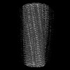
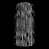
| Title | Cryo-EM structure of alpha-synuclein filaments doublet from Juvenile-onset synucleinopathy | |||||||||||||||
 Components Components |
| |||||||||||||||
 Keywords Keywords | PROTEIN FIBRIL / alpha-synuclein / amyloid / fibril / insertion / juvenile-onset synucleinopathy (JOS) / synucleinopathy | |||||||||||||||
| Function / homology |  Function and homology information Function and homology informationnegative regulation of mitochondrial electron transport, NADH to ubiquinone / neutral lipid metabolic process / regulation of acyl-CoA biosynthetic process / negative regulation of dopamine uptake involved in synaptic transmission / negative regulation of norepinephrine uptake / response to desipramine / positive regulation of SNARE complex assembly / positive regulation of hydrogen peroxide catabolic process / supramolecular fiber / regulation of synaptic vesicle recycling ...negative regulation of mitochondrial electron transport, NADH to ubiquinone / neutral lipid metabolic process / regulation of acyl-CoA biosynthetic process / negative regulation of dopamine uptake involved in synaptic transmission / negative regulation of norepinephrine uptake / response to desipramine / positive regulation of SNARE complex assembly / positive regulation of hydrogen peroxide catabolic process / supramolecular fiber / regulation of synaptic vesicle recycling / negative regulation of chaperone-mediated autophagy / regulation of reactive oxygen species biosynthetic process / positive regulation of protein localization to cell periphery / mitochondrial membrane organization / negative regulation of exocytosis / regulation of glutamate secretion / dopamine biosynthetic process / positive regulation of neurotransmitter secretion / response to iron(II) ion / regulation of macrophage activation / negative regulation of dopamine metabolic process / negative regulation of platelet-derived growth factor receptor signaling pathway / SNARE complex assembly / negative regulation of thrombin-activated receptor signaling pathway / Lewy body / regulation of locomotion / negative regulation of microtubule polymerization / synaptic vesicle priming / regulation of norepinephrine uptake / transporter regulator activity / protein kinase inhibitor activity / positive regulation of inositol phosphate biosynthetic process / synaptic vesicle transport / dopamine uptake involved in synaptic transmission / regulation of dopamine secretion / positive regulation of receptor recycling / cuprous ion binding / positive regulation of exocytosis / nuclear outer membrane / mitochondrial ATP synthesis coupled electron transport / dynein complex binding / synaptic vesicle exocytosis / response to magnesium ion / positive regulation of endocytosis / negative regulation of serotonin uptake / response to type II interferon / cysteine-type endopeptidase inhibitor activity / kinesin binding / regulation of presynapse assembly / synaptic vesicle endocytosis / alpha-tubulin binding / beta-tubulin binding / phospholipase binding / phospholipid metabolic process / supramolecular fiber organization / behavioral response to cocaine / cellular response to fibroblast growth factor stimulus / cellular response to epinephrine stimulus / inclusion body / Hsp70 protein binding / enzyme inhibitor activity / response to interleukin-1 / axon terminus / cellular response to copper ion / regulation of microtubule cytoskeleton organization / positive regulation of release of sequestered calcium ion into cytosol / SNARE binding / adult locomotory behavior / glutathione metabolic process / protein tetramerization / protein sequestering activity / phosphoprotein binding / tubulin binding / excitatory postsynaptic potential / microglial cell activation / ferrous iron binding / fatty acid metabolic process / phospholipid binding / PKR-mediated signaling / synapse organization / receptor internalization / regulation of long-term neuronal synaptic plasticity / protein destabilization / tau protein binding / enzyme activator activity / positive regulation of inflammatory response / terminal bouton / long-term synaptic potentiation / actin cytoskeleton / synaptic vesicle membrane / growth cone / actin binding / cellular response to oxidative stress / neuron apoptotic process / cell cortex / histone binding / response to lipopolysaccharide / microtubule binding / amyloid fibril formation / chemical synaptic transmission Similarity search - Function | |||||||||||||||
| Biological species |  Homo sapiens (human) Homo sapiens (human) | |||||||||||||||
| Method | ELECTRON MICROSCOPY / helical reconstruction / cryo EM / Resolution: 2.3 Å | |||||||||||||||
 Authors Authors | Yang, Y. / Garringer, J.H. / Shi, Y. / Lovestam, S. / Peak-Chew, S.Y. / Zhang, X.J. / Kotecha, A. / Bacioglu, M. / Koto, A. / Takao, M. ...Yang, Y. / Garringer, J.H. / Shi, Y. / Lovestam, S. / Peak-Chew, S.Y. / Zhang, X.J. / Kotecha, A. / Bacioglu, M. / Koto, A. / Takao, M. / Spillantini, G.M. / Ghetti, B. / Vidal, R. / Murzin, G.A. / Scheres, H.W.S. / Goedert, M. | |||||||||||||||
| Funding support |  United Kingdom, United Kingdom,  United States, 4items United States, 4items
| |||||||||||||||
 Citation Citation | Journal: Acta Neuropathol / Year: 2023 Title: New SNCA mutation and structures of α-synuclein filaments from juvenile-onset synucleinopathy. Authors: Yang Yang / Holly J Garringer / Yang Shi / Sofia Lövestam / Sew Peak-Chew / Xianjun Zhang / Abhay Kotecha / Mehtap Bacioglu / Atsuo Koto / Masaki Takao / Maria Grazia Spillantini / ...Authors: Yang Yang / Holly J Garringer / Yang Shi / Sofia Lövestam / Sew Peak-Chew / Xianjun Zhang / Abhay Kotecha / Mehtap Bacioglu / Atsuo Koto / Masaki Takao / Maria Grazia Spillantini / Bernardino Ghetti / Ruben Vidal / Alexey G Murzin / Sjors H W Scheres / Michel Goedert /    Abstract: A 21-nucleotide duplication in one allele of SNCA was identified in a previously described disease with abundant α-synuclein inclusions that we now call juvenile-onset synucleinopathy (JOS). This ...A 21-nucleotide duplication in one allele of SNCA was identified in a previously described disease with abundant α-synuclein inclusions that we now call juvenile-onset synucleinopathy (JOS). This mutation translates into the insertion of MAAAEKT after residue 22 of α-synuclein, resulting in a protein of 147 amino acids. Both wild-type and mutant proteins were present in sarkosyl-insoluble material that was extracted from frontal cortex of the individual with JOS and examined by electron cryo-microscopy. The structures of JOS filaments, comprising either a single protofilament, or a pair of protofilaments, revealed a new α-synuclein fold that differs from the folds of Lewy body diseases and multiple system atrophy (MSA). The JOS fold consists of a compact core, the sequence of which (residues 36-100 of wild-type α-synuclein) is unaffected by the mutation, and two disconnected density islands (A and B) of mixed sequences. There is a non-proteinaceous cofactor bound between the core and island A. The JOS fold resembles the common substructure of MSA Type I and Type II dimeric filaments, with its core segment approximating the C-terminal body of MSA protofilaments B and its islands mimicking the N-terminal arm of MSA protofilaments A. The partial similarity of JOS and MSA folds extends to the locations of their cofactor-binding sites. In vitro assembly of recombinant wild-type α-synuclein, its insertion mutant and their mixture yielded structures that were distinct from those of JOS filaments. Our findings provide insight into a possible mechanism of JOS fibrillation in which mutant α-synuclein of 147 amino acids forms a nucleus with the JOS fold, around which wild-type and mutant proteins assemble during elongation. | |||||||||||||||
| History |
|
- Structure visualization
| Structure viewer | Molecule: MolmilJmol/JSmol |
|---|
- Downloads & links
-Download
| PDBx/mmCIF format | 8bqw.cif.gz | 43 KB | Display | PDBx/mmCIF format |
|---|---|---|---|---|
| PDB format | pdb8bqw.ent.gz | 26.9 KB | Display | PDB format |
| PDBx/mmJSON format | 8bqw.json.gz | Tree view | PDBx/mmJSON format | |
| Others |  Other downloads Other downloads |
-Validation report
| Arichive directory | https://data.pdbj.org/pub/pdb/validation_reports/bq/8bqwftp://data.pdbj.org/pub/pdb/validation_reports/bq/8bqw | HTTPS FTP |
|---|
-Related structure data
| Related structure data | 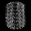 16189MC  8bqvC 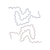 8ce7C  8cebC M: map data used to model this data C: citing same article ( |
|---|---|
| Similar structure data |


-Links
 PDBj
PDBj
- Assembly
| Deposited unit | 
| ||||||||||||||||
|---|---|---|---|---|---|---|---|---|---|---|---|---|---|---|---|---|---|
| 1 | x 5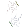
| ||||||||||||||||
| Noncrystallographic symmetry (NCS) | NCS oper:
|
-Components
| #1: Protein | Mass: 14476.108 Da / Num. of mol.: 2 / Source method: isolated from a natural source / Source: (natural) Homo sapiens (human) / References: UniProt: P37840#2: Protein/peptide | Mass: 783.958 Da / Num. of mol.: 2 / Source method: isolated from a natural source / Source: (natural) Homo sapiens (human)#3: Water | ChemComp-HOH / |  Mass: 18.015 Da / Num. of mol.: 9 / Source method: isolated from a natural source / Formula: H2O Mass: 18.015 Da / Num. of mol.: 9 / Source method: isolated from a natural source / Formula: H2O |
|---|
-Experimental details
-Experiment
| Experiment | Method: ELECTRON MICROSCOPY |
|---|---|
| EM experiment | Aggregation state: FILAMENT / 3D reconstruction method: helical reconstruction |
- Sample preparation
| Component | Name: Alpha-synuclein doublet filament extracted from the human brain with JOS Type: TISSUE / Entity ID: #1-#2 / Source: NATURAL |
|---|---|
| Source (natural) | Organism: Homo sapiens (human) |
| Buffer solution | pH: 7.5 |
| Specimen | Embedding applied: NO / Shadowing applied: NO / Staining applied: NO / Vitrification applied: YES |
| Vitrification | Cryogen name: ETHANE |
- Electron microscopy imaging
| Experimental equipment |  Model: Titan Krios / Image courtesy: FEI Company |
|---|---|
| Microscopy | Model: FEI TITAN KRIOS |
| Electron gun | Electron source:  FIELD EMISSION GUN / Accelerating voltage: 300 kV / Illumination mode: FLOOD BEAM FIELD EMISSION GUN / Accelerating voltage: 300 kV / Illumination mode: FLOOD BEAM |
| Electron lens | Mode: BRIGHT FIELD / Nominal defocus max: 1400 nm / Nominal defocus min: 600 nm |
| Image recording | Electron dose: 40 e/Å2 / Film or detector model: FEI FALCON IV (4k x 4k) |
- Processing
| Software | Name: REFMAC / Version: 5.8.0352 / Classification: refinement | ||||||||||||||||||||||||||||||||||||||||||||||||||||||||||||||||||||||||||||||||||||||||||||||||||||||||||
|---|---|---|---|---|---|---|---|---|---|---|---|---|---|---|---|---|---|---|---|---|---|---|---|---|---|---|---|---|---|---|---|---|---|---|---|---|---|---|---|---|---|---|---|---|---|---|---|---|---|---|---|---|---|---|---|---|---|---|---|---|---|---|---|---|---|---|---|---|---|---|---|---|---|---|---|---|---|---|---|---|---|---|---|---|---|---|---|---|---|---|---|---|---|---|---|---|---|---|---|---|---|---|---|---|---|---|---|
| EM software |
| ||||||||||||||||||||||||||||||||||||||||||||||||||||||||||||||||||||||||||||||||||||||||||||||||||||||||||
| CTF correction | Type: PHASE FLIPPING AND AMPLITUDE CORRECTION | ||||||||||||||||||||||||||||||||||||||||||||||||||||||||||||||||||||||||||||||||||||||||||||||||||||||||||
| Helical symmerty | Angular rotation/subunit: -1.11 ° / Axial rise/subunit: 4.71 Å / Axial symmetry: C2 | ||||||||||||||||||||||||||||||||||||||||||||||||||||||||||||||||||||||||||||||||||||||||||||||||||||||||||
| 3D reconstruction | Resolution: 2.3 Å / Resolution method: FSC 0.143 CUT-OFF / Num. of particles: 45038 / Symmetry type: HELICAL | ||||||||||||||||||||||||||||||||||||||||||||||||||||||||||||||||||||||||||||||||||||||||||||||||||||||||||
| Atomic model building | Details: servalcat | ||||||||||||||||||||||||||||||||||||||||||||||||||||||||||||||||||||||||||||||||||||||||||||||||||||||||||
| Refinement | Resolution: 2.3→149.76 Å / Cor.coef. Fo:Fc: 0.707 / SU B: 5.166 / SU ML: 0.121 / ESU R: 0.11 Stereochemistry target values: MAXIMUM LIKELIHOOD WITH PHASES Details: HYDROGENS HAVE BEEN ADDED IN THE RIDING POSITIONS
| ||||||||||||||||||||||||||||||||||||||||||||||||||||||||||||||||||||||||||||||||||||||||||||||||||||||||||
| Solvent computation | Solvent model: PARAMETERS FOR MASK CACLULATION | ||||||||||||||||||||||||||||||||||||||||||||||||||||||||||||||||||||||||||||||||||||||||||||||||||||||||||
| Displacement parameters | Biso mean: 52.265 Å2 | ||||||||||||||||||||||||||||||||||||||||||||||||||||||||||||||||||||||||||||||||||||||||||||||||||||||||||
| Refinement step | Cycle: 1 / Total: 1159 | ||||||||||||||||||||||||||||||||||||||||||||||||||||||||||||||||||||||||||||||||||||||||||||||||||||||||||
| Refine LS restraints |
|