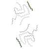
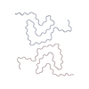
Journal: Acta Neuropathol / Year: 2023 Title: New SNCA mutation and structures of α-synuclein filaments from juvenile-onset synucleinopathy. Authors: Yang Yang / Holly J Garringer / Yang Shi / Sofia Lövestam / Sew Peak-Chew / Xianjun Zhang / Abhay Kotecha / Mehtap Bacioglu / Atsuo Koto / Masaki Takao / Maria Grazia Spillantini / ...Authors: Yang Yang / Holly J Garringer / Yang Shi / Sofia Lövestam / Sew Peak-Chew / Xianjun Zhang / Abhay Kotecha / Mehtap Bacioglu / Atsuo Koto / Masaki Takao / Maria Grazia Spillantini / Bernardino Ghetti / Ruben Vidal / Alexey G Murzin / Sjors H W Scheres / Michel Goedert / Abstract: A 21-nucleotide duplication in one allele of SNCA was identified in a previously described disease with abundant α-synuclein inclusions that we now call juvenile-onset synucleinopathy (JOS). This ...A 21-nucleotide duplication in one allele of SNCA was identified in a previously described disease with abundant α-synuclein inclusions that we now call juvenile-onset synucleinopathy (JOS). This mutation translates into the insertion of MAAAEKT after residue 22 of α-synuclein, resulting in a protein of 147 amino acids. Both wild-type and mutant proteins were present in sarkosyl-insoluble material that was extracted from frontal cortex of the individual with JOS and examined by electron cryo-microscopy. The structures of JOS filaments, comprising either a single protofilament, or a pair of protofilaments, revealed a new α-synuclein fold that differs from the folds of Lewy body diseases and multiple system atrophy (MSA). The JOS fold consists of a compact core, the sequence of which (residues 36-100 of wild-type α-synuclein) is unaffected by the mutation, and two disconnected density islands (A and B) of mixed sequences. There is a non-proteinaceous cofactor bound between the core and island A. The JOS fold resembles the common substructure of MSA Type I and Type II dimeric filaments, with its core segment approximating the C-terminal body of MSA protofilaments B and its islands mimicking the N-terminal arm of MSA protofilaments A. The partial similarity of JOS and MSA folds extends to the locations of their cofactor-binding sites. In vitro assembly of recombinant wild-type α-synuclein, its insertion mutant and their mixture yielded structures that were distinct from those of JOS filaments. Our findings provide insight into a possible mechanism of JOS fibrillation in which mutant α-synuclein of 147 amino acids forms a nucleus with the JOS fold, around which wild-type and mutant proteins assemble during elongation.
In the structure databanks used in Yorodumi, some data are registered as the other names, "COVID-19 virus" and "2019-nCoV". Here are the details of the virus and the list of structure data.
Jan 31, 2019. EMDB accession codes are about to change! (news from PDBe EMDB page)
EMDB accession codes are about to change! (news from PDBe EMDB page)
The allocation of 4 digits for EMDB accession codes will soon come to an end. Whilst these codes will remain in use, new EMDB accession codes will include an additional digit and will expand incrementally as the available range of codes is exhausted. The current 4-digit format prefixed with “EMD-” (i.e. EMD-XXXX) will advance to a 5-digit format (i.e. EMD-XXXXX), and so on. It is currently estimated that the 4-digit codes will be depleted around Spring 2019, at which point the 5-digit format will come into force.
The EM Navigator/Yorodumi systems omit the EMD- prefix.
Related info.:Q: What is EMD? / ID/Accession-code notation in Yorodumi/EM Navigator
Yorodumi is a browser for structure data from EMDB, PDB, SASBDB, etc.
This page is also the successor to EM Navigator detail page, and also detail information page/front-end page for Omokage search.
The word "yorodu" (or yorozu) is an old Japanese word meaning "ten thousand". "mi" (miru) is to see.
Related info.:EMDB / PDB / SASBDB / Comparison of 3 databanks / Yorodumi Search / Aug 31, 2016. New EM Navigator & Yorodumi / Yorodumi Papers / Jmol/JSmol / Function and homology information / Changes in new EM Navigator and Yorodumi
 Movie
Movie Controller


 Open data
 Basic information
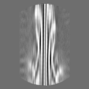
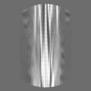
 Map data
 Sample
 Homo sapiens (human)
 Authors
 United Kingdom, 2 items
  Citation


 Structure visualization
 Downloads & links
 EMDB map data format
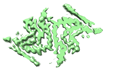 emd_16608.png
 http://ftp.pdbj.org/pub/emdb/structures/EMD-16608


 Z (Sec.)
 Y (Row.)
 X (Col.)


 Sample components
 Processing
 Electron microscopy
 FIELD EMISSION GUN

