| 登録情報 | データベース: PDB / ID: 8bo8
|
|---|
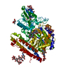
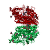
| タイトル | X-ray structure of human glutamate carboxypeptidase II (GCPII) in complex with an inhibitor P17 |
|---|
 要素 要素 | Glutamate carboxypeptidase 2 |
|---|
 キーワード キーワード | HYDROLASE / glutamate carboxypeptidase II (GCPII) / NAALADase / prostate-specific membrane antigen / inhibitor |
|---|
| 機能・相同性 |  機能・相同性情報 機能・相同性情報
Ac-Asp-Glu binding / tetrahydrofolyl-poly(glutamate) polymer binding / glutamate carboxypeptidase II / folic acid-containing compound metabolic process / C-terminal protein deglutamylation / Aspartate and asparagine metabolism / dipeptidase activity / carboxypeptidase activity / metallocarboxypeptidase activity / peptidase activity ...Ac-Asp-Glu binding / tetrahydrofolyl-poly(glutamate) polymer binding / glutamate carboxypeptidase II / folic acid-containing compound metabolic process / C-terminal protein deglutamylation / Aspartate and asparagine metabolism / dipeptidase activity / carboxypeptidase activity / metallocarboxypeptidase activity / peptidase activity / cell surface / proteolysis / extracellular exosome / metal ion binding / membrane / plasma membrane / cytoplasm類似検索 - 分子機能 Transferrin receptor-like, dimerisation domain / Transferrin receptor-like, dimerisation domain superfamily / Glutamate carboxypeptidase 2-like / Transferrin receptor-like dimerisation domain / PA domain superfamily / PA domain / PA domain / Peptidase M28 / Peptidase family M28類似検索 - ドメイン・相同性 |
|---|
| 生物種 |  Homo sapiens (ヒト) Homo sapiens (ヒト) |
|---|
| 手法 |  X線回折 / シンクロトロン / フーリエ合成 / 解像度: 1.55 Å X線回折 / シンクロトロン / フーリエ合成 / 解像度: 1.55 Å |
|---|
 データ登録者 データ登録者 | Motlova, L. / Barinka, C. / Benesova, M. |
|---|
| 資金援助 | 1件 |
|---|
 引用 引用 | ジャーナル: Acs Omega / 年: 2025
タイトル: Structure-Activity Relationships and Biological Insights into PSMA-617 and Its Derivatives with Modified Lipophilic Linker Regions.
著者: Schafer, M. / Bauder-Wust, U. / Roscher, M. / Motlova, L. / Kutilova, Z. / Remde, Y. / Klika, K.D. / Graf, J. / Barinka, C. / Benesova-Schafer, M. |
|---|
| 履歴 | | 登録 | 2022年11月15日 | 登録サイト: PDBE / 処理サイト: PDBE |
|---|
| 改定 1.0 | 2023年11月29日 | Provider: repository / タイプ: Initial release |
|---|
| 改定 1.1 | 2024年10月16日 | Group: Structure summary
カテゴリ: pdbx_entry_details / pdbx_modification_feature
Item: _pdbx_entry_details.has_protein_modification |
|---|
| 改定 1.2 | 2025年3月26日 | Group: Database references / カテゴリ: citation / citation_author
Item: _citation.country / _citation.journal_abbrev ..._citation.country / _citation.journal_abbrev / _citation.journal_id_CSD / _citation.journal_id_ISSN / _citation.journal_volume / _citation.page_first / _citation.page_last / _citation.pdbx_database_id_DOI / _citation.pdbx_database_id_PubMed / _citation.title / _citation.year |
|---|
|
|---|
 ムービー
ムービー コントローラー


 データを開く
 基本情報
基本情報 構造の表示
構造の表示 ダウンロードとリンク
 その他のダウンロード
 PDBj
PDBj
 集合体
集合体


 タイプ: D-saccharide, beta linking / 分子量: 221.208 Da / 分子数: 2 / 由来タイプ: 合成 / 式: C8H15NO6


 分子量: 65.409 Da / 分子数: 2 / 由来タイプ: 合成 / 式: Zn
 分子量: 40.078 Da / 分子数: 1 / 由来タイプ: 合成 / 式: Ca
 分子量: 35.453 Da / 分子数: 1 / 由来タイプ: 合成 / 式: Cl
 分子量: 22.990 Da / 分子数: 1 / 由来タイプ: 合成 / 式: Na
 分子量: 1018.117 Da / 分子数: 1 / 由来タイプ: 合成 / 式: C47H71N9O16 / タイプ: SUBJECT OF INVESTIGATION
 試料調製
試料調製 / ビームライン: 14.2 / 波長: 0.918 Å
 解析
解析