| Entry | Database: PDB / ID: 5o5t
|
|---|
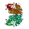
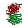
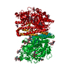
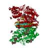
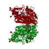
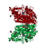
| Title | X-ray structure of human glutamate carboxypeptidase II (GCPII) in complex with a urea based inhibitor PSMA 1007 |
|---|
 Components Components | Glutamate carboxypeptidase 2 |
|---|
 Keywords Keywords | HYDROLASE / glutamate carboxypeptidase II (GCPII) / NAALADase / prostate-specific membrane antigen / urea based inhibitor |
|---|
| Function / homology |  Function and homology information Function and homology information
intestinal folate absorption / Ac-Asp-Glu binding / tetrahydrofolyl-poly(glutamate) polymer binding / glutamate carboxypeptidase II / Aspartate and asparagine metabolism / dipeptidase activity / : / positive regulation of glutamate receptor signaling pathway / carboxypeptidase activity / metallocarboxypeptidase activity ...intestinal folate absorption / Ac-Asp-Glu binding / tetrahydrofolyl-poly(glutamate) polymer binding / glutamate carboxypeptidase II / Aspartate and asparagine metabolism / dipeptidase activity / : / positive regulation of glutamate receptor signaling pathway / carboxypeptidase activity / metallocarboxypeptidase activity / peptidase activity / cell surface / proteolysis / extracellular exosome / membrane / metal ion binding / plasma membrane / cytoplasmSimilarity search - Function Transferrin receptor-like, dimerisation domain / Glucose Oxidase; domain 1 - #30 / Transferrin receptor-like, dimerisation domain / Transferrin receptor-like, dimerisation domain superfamily / Glutamate carboxypeptidase 2-like / Transferrin receptor-like dimerisation domain / PA domain / PA domain superfamily / Transcription Elongation Factor S-II; Chain A / PA domain ...Transferrin receptor-like, dimerisation domain / Glucose Oxidase; domain 1 - #30 / Transferrin receptor-like, dimerisation domain / Transferrin receptor-like, dimerisation domain superfamily / Glutamate carboxypeptidase 2-like / Transferrin receptor-like dimerisation domain / PA domain / PA domain superfamily / Transcription Elongation Factor S-II; Chain A / PA domain / Glucose Oxidase; domain 1 / Peptidase M28 / Peptidase family M28 / Zn peptidases / Aminopeptidase / 3-Layer(bba) Sandwich / Up-down Bundle / 3-Layer(aba) Sandwich / Mainly Alpha / Alpha BetaSimilarity search - Domain/homology |
|---|
| Biological species |  Homo sapiens (human) Homo sapiens (human) |
|---|
| Method |  X-RAY DIFFRACTION / SYNCHROTRON / MOLECULAR REPLACEMENT / Resolution: 1.43 Å X-RAY DIFFRACTION / SYNCHROTRON / MOLECULAR REPLACEMENT / Resolution: 1.43 Å |
|---|
 Authors Authors | Barinka, C. / Novakova, Z. / Motlova, L. |
|---|
 Citation Citation | Journal: To Be Published
Title: X-ray structure of human glutamate carboxypeptidase II (GCPII) in complex with a urea based inhibitor PSMA 1007
Authors: Barinka, C. / Novakova, Z. / Motlova, L. |
|---|
| History | | Deposition | Jun 2, 2017 | Deposition site: PDBE / Processing site: PDBE |
|---|
| Revision 1.0 | Jun 13, 2018 | Provider: repository / Type: Initial release |
|---|
| Revision 2.0 | Jul 29, 2020 | Group: Advisory / Atomic model ...Advisory / Atomic model / Data collection / Derived calculations / Structure summary
Category: atom_site / atom_site_anisotrop ...atom_site / atom_site_anisotrop / chem_comp / entity / pdbx_branch_scheme / pdbx_chem_comp_identifier / pdbx_entity_branch / pdbx_entity_branch_descriptor / pdbx_entity_branch_link / pdbx_entity_branch_list / pdbx_entity_nonpoly / pdbx_nonpoly_scheme / pdbx_struct_assembly_gen / pdbx_struct_conn_angle / pdbx_validate_symm_contact / struct_asym / struct_conn / struct_site / struct_site_gen
Item: _atom_site.B_iso_or_equiv / _atom_site.Cartn_x ..._atom_site.B_iso_or_equiv / _atom_site.Cartn_x / _atom_site.Cartn_y / _atom_site.Cartn_z / _atom_site.auth_asym_id / _atom_site.auth_atom_id / _atom_site.auth_comp_id / _atom_site.auth_seq_id / _atom_site.label_asym_id / _atom_site.label_atom_id / _atom_site.label_comp_id / _atom_site.label_entity_id / _atom_site.type_symbol / _atom_site_anisotrop.U[1][1] / _atom_site_anisotrop.U[1][2] / _atom_site_anisotrop.U[1][3] / _atom_site_anisotrop.U[2][2] / _atom_site_anisotrop.U[2][3] / _atom_site_anisotrop.U[3][3] / _atom_site_anisotrop.pdbx_auth_asym_id / _atom_site_anisotrop.pdbx_auth_atom_id / _atom_site_anisotrop.pdbx_auth_comp_id / _atom_site_anisotrop.pdbx_auth_seq_id / _atom_site_anisotrop.pdbx_label_asym_id / _atom_site_anisotrop.pdbx_label_atom_id / _atom_site_anisotrop.pdbx_label_comp_id / _atom_site_anisotrop.type_symbol / _chem_comp.name / _chem_comp.type / _pdbx_struct_assembly_gen.asym_id_list / _pdbx_struct_conn_angle.ptnr1_auth_comp_id / _pdbx_struct_conn_angle.ptnr1_auth_seq_id / _pdbx_struct_conn_angle.ptnr1_label_asym_id / _pdbx_struct_conn_angle.ptnr1_label_atom_id / _pdbx_struct_conn_angle.ptnr1_label_comp_id / _pdbx_struct_conn_angle.ptnr2_label_asym_id / _pdbx_struct_conn_angle.ptnr3_auth_comp_id / _pdbx_struct_conn_angle.ptnr3_auth_seq_id / _pdbx_struct_conn_angle.ptnr3_label_asym_id / _pdbx_struct_conn_angle.ptnr3_label_atom_id / _pdbx_struct_conn_angle.ptnr3_label_comp_id / _pdbx_struct_conn_angle.value / _pdbx_validate_symm_contact.auth_asym_id_1 / _pdbx_validate_symm_contact.auth_seq_id_1 / _struct_conn.conn_type_id / _struct_conn.id / _struct_conn.pdbx_dist_value / _struct_conn.pdbx_leaving_atom_flag / _struct_conn.pdbx_role / _struct_conn.ptnr1_auth_asym_id / _struct_conn.ptnr1_auth_comp_id / _struct_conn.ptnr1_auth_seq_id / _struct_conn.ptnr1_label_asym_id / _struct_conn.ptnr1_label_atom_id / _struct_conn.ptnr1_label_comp_id / _struct_conn.ptnr1_label_seq_id / _struct_conn.ptnr2_auth_asym_id / _struct_conn.ptnr2_auth_comp_id / _struct_conn.ptnr2_auth_seq_id / _struct_conn.ptnr2_label_asym_id / _struct_conn.ptnr2_label_atom_id / _struct_conn.ptnr2_label_comp_id
Description: Carbohydrate remediation / Provider: repository / Type: Remediation |
|---|
| Revision 2.1 | Oct 23, 2024 | Group: Data collection / Database references ...Data collection / Database references / Derived calculations / Structure summary
Category: chem_comp / chem_comp_atom ...chem_comp / chem_comp_atom / chem_comp_bond / database_2 / pdbx_entry_details / pdbx_modification_feature / struct_conn
Item: _chem_comp.pdbx_synonyms / _database_2.pdbx_DOI ..._chem_comp.pdbx_synonyms / _database_2.pdbx_DOI / _database_2.pdbx_database_accession / _struct_conn.pdbx_leaving_atom_flag |
|---|
|
|---|
 Movie
Movie Controller

 Yorodumi
 Open data
 Basic information
 Structure visualization
 Downloads & links
 Other downloads


 PDBj
PDBj
 Assembly


 Type: D-saccharide, beta linking / Mass: 221.208 Da / Num. of mol.: 4


 Mass: 65.409 Da / Num. of mol.: 2 / Source method: obtained synthetically / Formula: Zn
 Mass: 40.078 Da / Num. of mol.: 1 / Source method: obtained synthetically / Formula: Ca
 Mass: 35.453 Da / Num. of mol.: 1 / Source method: obtained synthetically / Formula: Cl
 Mass: 1031.003 Da / Num. of mol.: 1 / Source method: obtained synthetically / Formula: C49H55FN8O16
 Sample preparation
 / Beamline: P13 (MX1) / Wavelength: 0.9796 Å
 Processing