 Movie
Movie Controller

[English] 日本語
 Yorodumi
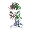
- PDB-8be1: SARS-CoV-2 RBD in complex with a Fab fragment of a neutralising a... -
+ Open data

- Basic information
| Entry | Database: PDB / ID: 8be1 | ||||||
|---|---|---|---|---|---|---|---|
| Title | SARS-CoV-2 RBD in complex with a Fab fragment of a neutralising antibody mRBD2 | ||||||
 Components Components |
| ||||||
 Keywords Keywords | IMMUNE SYSTEM / FAB / antibody / SARS-COV-2 / RBD / neutralising | ||||||
| Function / homology |  Function and homology information Function and homology informationMaturation of spike protein / viral translation / Translation of Structural Proteins / Virion Assembly and Release / host cell surface / host extracellular space / suppression by virus of host tetherin activity / Induction of Cell-Cell Fusion / structural constituent of virion / entry receptor-mediated virion attachment to host cell ...Maturation of spike protein / viral translation / Translation of Structural Proteins / Virion Assembly and Release / host cell surface / host extracellular space / suppression by virus of host tetherin activity / Induction of Cell-Cell Fusion / structural constituent of virion / entry receptor-mediated virion attachment to host cell / host cell endoplasmic reticulum-Golgi intermediate compartment membrane / membrane fusion / receptor-mediated endocytosis of virus by host cell / Attachment and Entry / positive regulation of viral entry into host cell / receptor-mediated virion attachment to host cell / receptor ligand activity / symbiont-mediated suppression of host innate immune response / host cell surface receptor binding / fusion of virus membrane with host plasma membrane / fusion of virus membrane with host endosome membrane / viral envelope / virion attachment to host cell / SARS-CoV-2 activates/modulates innate and adaptive immune responses / host cell plasma membrane / virion membrane / identical protein binding / membrane / plasma membrane Similarity search - Function | ||||||
| Biological species |    Severe acute respiratory syndrome coronavirus 2 Severe acute respiratory syndrome coronavirus 2 | ||||||
| Method |  X-RAY DIFFRACTION / SYNCHROTRON / MOLECULAR REPLACEMENT / Resolution: 1.98 Å X-RAY DIFFRACTION / SYNCHROTRON / MOLECULAR REPLACEMENT / Resolution: 1.98 Å | ||||||
 Authors Authors | Lulla, A. / Brear, P. / Fischer, K. / Hollfelder, F. / Hyvonen, M. | ||||||
| Funding support | 1items
| ||||||
 Citation Citation | Journal: Biorxiv / Year: 2023 Title: Microfluidics-enabled fluorescence-activated cell sorting of single pathogen-specific antibody secreting cells for the rapid discovery of monoclonal antibodies Authors: Fischer, K. / Lulla, A. / So, T.Y. / Pereyra-Gerber, P. / Raybould, M.I.J. / Kohler, T.N. / Kaminski, T.S. / Yam-Puc, J.C. / Hughes, R. / Leiss-Maier, F. / Brear, P. / Matheson, N.J. / ...Authors: Fischer, K. / Lulla, A. / So, T.Y. / Pereyra-Gerber, P. / Raybould, M.I.J. / Kohler, T.N. / Kaminski, T.S. / Yam-Puc, J.C. / Hughes, R. / Leiss-Maier, F. / Brear, P. / Matheson, N.J. / Deane, C.M. / Hyvonen, M. / Thaventhiran, J.E.D. / Hollfelder, F. | ||||||
| History |
|
- Structure visualization
| Structure viewer | Molecule: MolmilJmol/JSmol |
|---|
- Downloads & links
-Download
| PDBx/mmCIF format | 8be1.cif.gz | 254.3 KB | Display | PDBx/mmCIF format |
|---|---|---|---|---|
| PDB format | pdb8be1.ent.gz | 201.7 KB | Display | PDB format |
| PDBx/mmJSON format | 8be1.json.gz | Tree view | PDBx/mmJSON format | |
| Others |  Other downloads Other downloads |
-Validation report
| Summary document | 8be1_validation.pdf.gz | 493.5 KB | Display | wwPDB validaton report |
|---|---|---|---|---|
| Full document | 8be1_full_validation.pdf.gz | 509.6 KB | Display | |
| Data in XML | 8be1_validation.xml.gz | 46 KB | Display | |
| Data in CIF | 8be1_validation.cif.gz | 64.4 KB | Display | |
| Arichive directory | https://data.pdbj.org/pub/pdb/validation_reports/be/8be1ftp://data.pdbj.org/pub/pdb/validation_reports/be/8be1 | HTTPS FTP |
-Related structure data

-Links
 PDBj
PDBj


- Assembly
| Deposited unit | 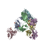
| ||||||||||
|---|---|---|---|---|---|---|---|---|---|---|---|
| 1 | 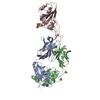
| ||||||||||
| 2 | 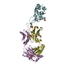
| ||||||||||
| Unit cell |
|
-Components
| #1: Antibody | Mass: 24907.758 Da / Num. of mol.: 2 Source method: isolated from a genetically manipulated source Source: (gene. exp.)  #2: Antibody | Mass: 23851.346 Da / Num. of mol.: 2 Source method: isolated from a genetically manipulated source Source: (gene. exp.) #3: Protein | Mass: 22074.678 Da / Num. of mol.: 2 Source method: isolated from a genetically manipulated source Source: (gene. exp.) Severe acute respiratory syndrome coronavirus 2Gene: S, 2 / Plasmid: pExp-Zbasic / Production host: #4: Chemical | ChemComp-SO4 /   Mass: 96.063 Da / Num. of mol.: 17 / Source method: obtained synthetically / Formula: SO4 Mass: 96.063 Da / Num. of mol.: 17 / Source method: obtained synthetically / Formula: SO4#5: Water | ChemComp-HOH / |  Mass: 18.015 Da / Num. of mol.: 286 / Source method: isolated from a natural source / Formula: H2O Mass: 18.015 Da / Num. of mol.: 286 / Source method: isolated from a natural source / Formula: H2OHas ligand of interest | N | |
|---|
-Experimental details
-Experiment
| Experiment | Method: X-RAY DIFFRACTION / Number of used crystals: 1 |
|---|
- Sample preparation
| Crystal | Density Matthews: 2.47 Å3/Da / Density % sol: 50.29 % |
|---|---|
| Crystal grow | Temperature: 281 K / Method: vapor diffusion, sitting drop / pH: 6.5 Details: 0.1 M Bis Tris Propane pH 6.5 0.2 M Potassium thiocyanate 20 % w/v PEG 3350 10 % v/v Ethylene glycol |
-Data collection
| Diffraction | Mean temperature: 100 K / Serial crystal experiment: N | ||||||||||||||||||||||||||||||
|---|---|---|---|---|---|---|---|---|---|---|---|---|---|---|---|---|---|---|---|---|---|---|---|---|---|---|---|---|---|---|---|
| Diffraction source | Source: SYNCHROTRON / Site: Diamond  / Beamline: I04 / Wavelength: 0.9795 Å / Beamline: I04 / Wavelength: 0.9795 Å | ||||||||||||||||||||||||||||||
| Detector | Type: DECTRIS EIGER X 16M / Detector: PIXEL / Date: Oct 12, 2022 | ||||||||||||||||||||||||||||||
| Radiation | Protocol: SINGLE WAVELENGTH / Monochromatic (M) / Laue (L): M / Scattering type: x-ray | ||||||||||||||||||||||||||||||
| Radiation wavelength | Wavelength: 0.9795 Å / Relative weight: 1 | ||||||||||||||||||||||||||||||
| Reflection | Resolution: 1.98→52.05 Å / Num. obs: 95377 / % possible obs: 100 % / Redundancy: 17.5 % / Biso Wilson estimate: 59.19 Å2 / CC1/2: 0.999 / Rmerge(I) obs: 0.109 / Rpim(I) all: 0.027 / Rrim(I) all: 0.112 / Net I/σ(I): 13.2 / Num. measured all: 1669839 / Scaling rejects: 3 | ||||||||||||||||||||||||||||||
| Reflection shell | Diffraction-ID: 1
|
- Processing
| Software |
| ||||||||||||||||||||||||||||||||||||||||||||||||||||||||||||
|---|---|---|---|---|---|---|---|---|---|---|---|---|---|---|---|---|---|---|---|---|---|---|---|---|---|---|---|---|---|---|---|---|---|---|---|---|---|---|---|---|---|---|---|---|---|---|---|---|---|---|---|---|---|---|---|---|---|---|---|---|---|
| Refinement | Method to determine structure: MOLECULAR REPLACEMENT Starting model: 3BGF, 6YM0 Resolution: 1.98→39.02 Å / Cor.coef. Fo:Fc: 0.938 / Cor.coef. Fo:Fc free: 0.923 / SU R Cruickshank DPI: 0.21 / Cross valid method: THROUGHOUT / SU R Blow DPI: 0.202 / SU Rfree Blow DPI: 0.178 / SU Rfree Cruickshank DPI: 0.183
| ||||||||||||||||||||||||||||||||||||||||||||||||||||||||||||
| Displacement parameters | Biso mean: 64.95 Å2
| ||||||||||||||||||||||||||||||||||||||||||||||||||||||||||||
| Refine analyze | Luzzati coordinate error obs: 0.35 Å | ||||||||||||||||||||||||||||||||||||||||||||||||||||||||||||
| Refinement step | Cycle: LAST / Resolution: 1.98→39.02 Å
| ||||||||||||||||||||||||||||||||||||||||||||||||||||||||||||
| Refine LS restraints |
| ||||||||||||||||||||||||||||||||||||||||||||||||||||||||||||
| LS refinement shell | Resolution: 1.98→2 Å
|
