 Movie
Movie Controller

[English] 日本語
 Yorodumi
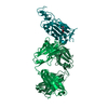
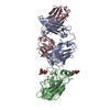
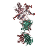
- PDB-6ym0: Crystal structure of the SARS-CoV-2 receptor binding domain in co... -
+ Open data

- Basic information
| Entry | Database: PDB / ID: 6ym0 | ||||||
|---|---|---|---|---|---|---|---|
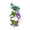
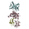
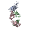
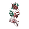
| Title | Crystal structure of the SARS-CoV-2 receptor binding domain in complex with CR3022 Fab (crystal form 1) | ||||||
 Components Components |
| ||||||
 Keywords Keywords | VIRAL PROTEIN / SARS-CoV-2 / receptor binding domain (RBD) / CR3022 / Vagabond | ||||||
| Function / homology |  Function and homology information Function and homology informationsymbiont-mediated disruption of host tissue / Maturation of spike protein / Translation of Structural Proteins / Virion Assembly and Release / host cell surface / host extracellular region / symbiont-mediated-mediated suppression of host tetherin activity / Induction of Cell-Cell Fusion / structural constituent of virion / positive regulation of viral entry into host cell ...symbiont-mediated disruption of host tissue / Maturation of spike protein / Translation of Structural Proteins / Virion Assembly and Release / host cell surface / host extracellular region / symbiont-mediated-mediated suppression of host tetherin activity / Induction of Cell-Cell Fusion / structural constituent of virion / positive regulation of viral entry into host cell / membrane fusion / host cell endoplasmic reticulum-Golgi intermediate compartment membrane / Attachment and Entry / entry receptor-mediated virion attachment to host cell / receptor-mediated virion attachment to host cell / host cell surface receptor binding / symbiont-mediated suppression of host innate immune response / endocytosis involved in viral entry into host cell / receptor ligand activity / fusion of virus membrane with host plasma membrane / fusion of virus membrane with host endosome membrane / viral envelope / symbiont entry into host cell / virion attachment to host cell / host cell plasma membrane / SARS-CoV-2 activates/modulates innate and adaptive immune responses / virion membrane / membrane / identical protein binding / plasma membrane Similarity search - Function | ||||||
| Biological species |   Severe acute respiratory syndrome coronavirus 2 Severe acute respiratory syndrome coronavirus 2 Homo sapiens (human) Homo sapiens (human) | ||||||
| Method |  X-RAY DIFFRACTION / SYNCHROTRON / MOLECULAR REPLACEMENT / Resolution: 4.36 Å X-RAY DIFFRACTION / SYNCHROTRON / MOLECULAR REPLACEMENT / Resolution: 4.36 Å | ||||||
 Authors Authors | Huo, J. / Zhao, Y. / Ren, J. / Zhou, D. / Ginn, H.M. / Fry, E.E. / Owens, R. / Stuart, D.I. | ||||||
| Funding support |  United Kingdom, 1items United Kingdom, 1items
| ||||||
 Citation Citation | Journal: Cell Host Microbe / Year: 2020 Title: Neutralization of SARS-CoV-2 by Destruction of the Prefusion Spike. Authors: Jiandong Huo / Yuguang Zhao / Jingshan Ren / Daming Zhou / Helen M E Duyvesteyn / Helen M Ginn / Loic Carrique / Tomas Malinauskas / Reinis R Ruza / Pranav N M Shah / Tiong Kit Tan / Pramila ...Authors: Jiandong Huo / Yuguang Zhao / Jingshan Ren / Daming Zhou / Helen M E Duyvesteyn / Helen M Ginn / Loic Carrique / Tomas Malinauskas / Reinis R Ruza / Pranav N M Shah / Tiong Kit Tan / Pramila Rijal / Naomi Coombes / Kevin R Bewley / Julia A Tree / Julika Radecke / Neil G Paterson / Piyada Supasa / Juthathip Mongkolsapaya / Gavin R Screaton / Miles Carroll / Alain Townsend / Elizabeth E Fry / Raymond J Owens / David I Stuart /  Abstract: There are as yet no licensed therapeutics for the COVID-19 pandemic. The causal coronavirus (SARS-CoV-2) binds host cells via a trimeric spike whose receptor binding domain (RBD) recognizes ...There are as yet no licensed therapeutics for the COVID-19 pandemic. The causal coronavirus (SARS-CoV-2) binds host cells via a trimeric spike whose receptor binding domain (RBD) recognizes angiotensin-converting enzyme 2, initiating conformational changes that drive membrane fusion. We find that the monoclonal antibody CR3022 binds the RBD tightly, neutralizing SARS-CoV-2, and report the crystal structure at 2.4 Å of the Fab/RBD complex. Some crystals are suitable for screening for entry-blocking inhibitors. The highly conserved, structure-stabilizing CR3022 epitope is inaccessible in the prefusion spike, suggesting that CR3022 binding facilitates conversion to the fusion-incompetent post-fusion state. Cryogenic electron microscopy (cryo-EM) analysis confirms that incubation of spike with CR3022 Fab leads to destruction of the prefusion trimer. Presentation of this cryptic epitope in an RBD-based vaccine might advantageously focus immune responses. Binders at this epitope could be useful therapeutically, possibly in synergy with an antibody that blocks receptor attachment. | ||||||
| History |
|
- Structure visualization
| Structure viewer | Molecule: MolmilJmol/JSmol |
|---|
- Downloads & links
-Download
| PDBx/mmCIF format | 6ym0.cif.gz | 120.9 KB | Display | PDBx/mmCIF format |
|---|---|---|---|---|
| PDB format | pdb6ym0.ent.gz | 90 KB | Display | PDB format |
| PDBx/mmJSON format | 6ym0.json.gz | Tree view | PDBx/mmJSON format | |
| Others |  Other downloads Other downloads |
-Validation report
| Arichive directory | https://data.pdbj.org/pub/pdb/validation_reports/ym/6ym0ftp://data.pdbj.org/pub/pdb/validation_reports/ym/6ym0 | HTTPS FTP |
|---|
-Related structure data
| Related structure data | 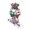 6ylaC 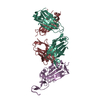 6yorC 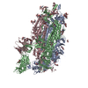 6z97C  4kmtS 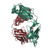 5i1dS 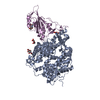 6m0jS C: citing same article ( S: Starting model for refinement |
|---|---|
| Similar structure data |


-Links
 PDBj
PDBj


- Assembly
| Deposited unit | 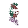
| ||||||||
|---|---|---|---|---|---|---|---|---|---|
| 1 |
| ||||||||
| Unit cell |
|
-Components
| #1: Protein | Mass: 24003.848 Da / Num. of mol.: 1 Source method: isolated from a genetically manipulated source Source: (gene. exp.) Severe acute respiratory syndrome coronavirus 2Gene: S, 2 / Production host: Homo sapiens (human) / References: UniProt: P0DTC2 |
|---|---|
| #2: Protein | Mass: 24354.412 Da / Num. of mol.: 1 Source method: isolated from a genetically manipulated source Source: (gene. exp.) Homo sapiens (human) / Production host: Homo sapiens (human) |
| #3: Antibody | Mass: 24289.885 Da / Num. of mol.: 1 Source method: isolated from a genetically manipulated source Source: (gene. exp.) Homo sapiens (human) / Production host: Homo sapiens (human) |
| Has protein modification | Y |
-Experimental details
-Experiment
| Experiment | Method: X-RAY DIFFRACTION / Number of used crystals: 1 |
|---|
- Sample preparation
| Crystal grow | Temperature: 293 K / Method: vapor diffusion, sitting drop / pH: 8 Details: 0.1 M Sodium malonate, 0.1 M Tris pH 8.0 and 30% w/v Polyethylene glycol 1,000. |
|---|
-Data collection
| Diffraction | Mean temperature: 100 K / Serial crystal experiment: N |
|---|---|
| Diffraction source | Source: SYNCHROTRON / Site: Diamond / Beamline: I03 / Wavelength: 0.9763 Å |
| Detector | Type: DECTRIS EIGER2 XE 16M / Detector: PIXEL / Date: Mar 6, 2020 |
| Radiation | Protocol: SINGLE WAVELENGTH / Monochromatic (M) / Laue (L): M / Scattering type: x-ray |
| Radiation wavelength | Wavelength: 0.9763 Å / Relative weight: 1 |
| Reflection | Resolution: 4.36→80.5 Å / Num. obs: 18822 / % possible obs: 100 % / Redundancy: 51.5 % / CC1/2: 0.952 / Rmerge(I) obs: 0.683 / Rpim(I) all: 0.097 / Net I/σ(I): 4 |
| Reflection shell | Resolution: 4.36→4.44 Å / Num. unique obs: 931 / CC1/2: 0.316 / % possible all: 100 |
- Processing
| Software |
| ||||||||||||||||||
|---|---|---|---|---|---|---|---|---|---|---|---|---|---|---|---|---|---|---|---|
| Refinement | Method to determine structure: MOLECULAR REPLACEMENT Starting model: 6M0J, 4KMT, 5I1D Resolution: 4.36→35 Å / Cross valid method: FREE R-VALUE
| ||||||||||||||||||
| Refinement step | Cycle: LAST / Resolution: 4.36→35 Å
|
