 ムービー
ムービー コントローラー


+ データを開く

- 基本情報
基本情報
| 登録情報 | データベース: PDB / ID: 8bcu | |||||||||
|---|---|---|---|---|---|---|---|---|---|---|
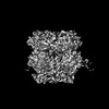
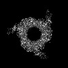
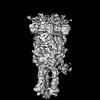
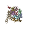
| タイトル | Cryo-EM structure of the proximal end of bacteriophage T5 tail, after interaction with its receptor : p142 tail terminator protein hexamer and pb6 tail tube protein trimer | |||||||||
 要素 要素 |
| |||||||||
 キーワード キーワード | VIRAL PROTEIN / Bacteriophage / Siphophage / T5 / tail terminator / Trp | |||||||||
| 機能・相同性 |  機能・相同性情報 機能・相同性情報virus tail, tube / symbiont genome ejection through host cell envelope, long flexible tail mechanism / viral tail assembly / virus tail / viral release from host cell by cytolysis 類似検索 - 分子機能 | |||||||||
| 生物種 |  Escherichia phage T5 (ファージ) Escherichia phage T5 (ファージ) | |||||||||
| 手法 | 電子顕微鏡法 / 単粒子再構成法 / クライオ電子顕微鏡法 / 解像度: 4.05 Å | |||||||||
 データ登録者 データ登録者 | Linares, R. / Effantin, G. / Breyton, C. | |||||||||
| 資金援助 |  フランス, 2件 フランス, 2件
| |||||||||
 引用 引用 | ジャーナル: J Virol / 年: 2025 タイトル: About bacteriophage tail terminator and tail completion proteins: structure of the proximal extremity of siphophage T5 tail. 著者: Romain Linares / Cécile Breyton / 要旨: Bacteriophages are viruses infecting bacteria. The vast majority of them bear a tail, allowing host recognition, cell wall perforation, and DNA injection into the host cytoplasm. Using electron cryo- ...Bacteriophages are viruses infecting bacteria. The vast majority of them bear a tail, allowing host recognition, cell wall perforation, and DNA injection into the host cytoplasm. Using electron cryo-microscopy (cryo-EM) and single particle analysis, we determined the organization of the tail proximal extremity of siphophage T5 that possesses a long flexible tail and solved the structure of its tail terminator protein p142 (TrP). It allowed us to confirm the common evolutionary origin between T5 TrP and other known or putative TrPs from siphophages, myophages, and bacterial tail-like machines, despite very poor sequence conservation. By also determining the structure of the T5 tail proximal extremity after interaction with T5 bacterial receptor FhuA, we showed that no conformational changes occur in TrP and confirmed that the infection signal transduction is not carried by the tube itself. We also investigated the location of T5 Neck1 or tail completion protein p143 (TCP) and showed, thanks to a combination of cryo-EM and structure prediction using Alphafold2, that it is not located at the capsid-to-tail interface as suggested by its position in the genome, but instead, very unexpectedly, on the side of T5 tail tip, and that it appears to be monomeric. Based on structure comparison with other putative TCPs predicted structures, this feature could not be shared by other TCPs and questions the affiliation of p143 to this family of protein.IMPORTANCEBacteriophages, viruses infecting bacteria, are the most abundant living entities on Earth. They are present in all ecosystems where bacteria develop and are instrumental in the regulation, diversity, evolution, and pathogeny of microbial populations. Moreover, with the increasing number of pathogenic strains resistant to antibiotics, virulent phages are considered a serious alternative or complement to classical treatments. 96% of all phages present a tail that allows host recognition and safe channeling of the DNA to the host cytoplasm. We present the atomic model of the proximal extremity of the siphophage T5 tail, confirming structural similarities with other phages. This structure, combined with results previously published and further explored, also allowed a review and a discussion on the role and localization of a mysterious tail protein, the tail completion protein, which is known to be present in the phage tails, but that was never identified in a phage structure. | |||||||||
| 履歴 |
|
- 構造の表示
構造の表示
| 構造ビューア | 分子: MolmilJmol/JSmol |
|---|
- ダウンロードとリンク
-ダウンロード
| PDBx/mmCIF形式 | 8bcu.cif.gz | 398.8 KB | 表示 | PDBx/mmCIF形式 |
|---|---|---|---|---|
| PDB形式 | pdb8bcu.ent.gz | 表示 | PDB形式 | |
| PDBx/mmJSON形式 | 8bcu.json.gz | ツリー表示 | PDBx/mmJSON形式 | |
| その他 |  その他のダウンロード その他のダウンロード |
-検証レポート
| アーカイブディレクトリ | https://data.pdbj.org/pub/pdb/validation_reports/bc/8bcuftp://data.pdbj.org/pub/pdb/validation_reports/bc/8bcu | HTTPS FTP |
|---|
-関連構造データ
-リンク
 PDBj
PDBj- 集合体
集合体
| 登録構造単位 | 
|
|---|---|
| 1 |
|
-要素
| #1: タンパク質 | 分子量: 18378.643 Da / 分子数: 6 / 由来タイプ: 天然 / 由来: (天然) Escherichia phage T5 (ファージ) / 参照: UniProt: Q6QGE1#2: タンパク質 | 分子量: 50459.215 Da / 分子数: 3 / 由来タイプ: 天然 / 由来: (天然) Escherichia phage T5 (ファージ) / 参照: UniProt: Q6QGE2Has protein modification | N | |
|---|
-実験情報
-実験
| 実験 | 手法: 電子顕微鏡法 |
|---|---|
| EM実験 | 試料の集合状態: PARTICLE / 3次元再構成法: 単粒子再構成法 |
- 試料調製
試料調製
| 構成要素 | 名称: Escherichia virus T5 / タイプ: VIRUS 詳細: Pure T5 tails obtained by infecting E. coli F strain with the amber mutant phage T5D20am30d, incubated with the bacterial receptor FhuA reconstituted in nanodiscs Entity ID: all / 由来: NATURAL | |||||||||||||||||||||||||
|---|---|---|---|---|---|---|---|---|---|---|---|---|---|---|---|---|---|---|---|---|---|---|---|---|---|---|
| 分子量 | 実験値: NO | |||||||||||||||||||||||||
| 由来(天然) | 生物種: Escherichia virus T5 (ウイルス) / 株: T5D20am30d | |||||||||||||||||||||||||
| ウイルスについての詳細 | 中空か: NO / エンベロープを持つか: NO / 単離: STRAIN / タイプ: VIRUS-LIKE PARTICLE | |||||||||||||||||||||||||
| 天然宿主 | 生物種: Escherichia coli / 株: F | |||||||||||||||||||||||||
| 緩衝液 | pH: 8 | |||||||||||||||||||||||||
| 緩衝液成分 |
| |||||||||||||||||||||||||
| 試料 | 包埋: NO / シャドウイング: NO / 染色: NO / 凍結: YES 詳細: Pure T5 tails obtained by infecting E. coli F strain with the amber mutant phage T5D20am30d, incubated with the bacterial receptor FhuA reconstituted in nanodiscs | |||||||||||||||||||||||||
| 試料支持 | 詳細: 25 mA / グリッドの材料: COPPER/RHODIUM / グリッドのサイズ: 300 divisions/in. / グリッドのタイプ: Quantifoil R2/1 | |||||||||||||||||||||||||
| 急速凍結 | 装置: FEI VITROBOT MARK IV / 凍結剤: ETHANE / 湿度: 100 % / 凍結前の試料温度: 293.15 K 詳細: 3 uL of T5 tails + FhuA-nanodisc sample were deposited on a freshly glow discharged EM grid and plunge-frozen in nitrogen-cooled liquid ethane using a ThermoFisher Mark IV Vitrobot device ...詳細: 3 uL of T5 tails + FhuA-nanodisc sample were deposited on a freshly glow discharged EM grid and plunge-frozen in nitrogen-cooled liquid ethane using a ThermoFisher Mark IV Vitrobot device (100 percent humidity, 20 Celsius degrees, 5 s blotting time, blot force 0) |
- 電子顕微鏡撮影
| 実験機器 |  モデル: Titan Krios / 画像提供: FEI Company |
|---|---|
| 顕微鏡 | モデル: FEI TITAN KRIOS |
| 電子銃 | 電子線源:  FIELD EMISSION GUN / 加速電圧: 300 kV / 照射モード: FLOOD BEAM FIELD EMISSION GUN / 加速電圧: 300 kV / 照射モード: FLOOD BEAM |
| 電子レンズ | モード: BRIGHT FIELD / 倍率(公称値): 105000 X / 最大 デフォーカス(公称値): 3000 nm / 最小 デフォーカス(公称値): 1000 nm / Cs: 2.7 mm |
| 試料ホルダ | 凍結剤: NITROGEN 試料ホルダーモデル: FEI TITAN KRIOS AUTOGRID HOLDER |
| 撮影 | 電子線照射量: 40 e/Å2 / 検出モード: COUNTING フィルム・検出器のモデル: GATAN K2 SUMMIT (4k x 4k) |
| 画像スキャン | 動画フレーム数/画像: 40 |
- 解析
解析
| ソフトウェア | 名称: PHENIX / バージョン: 1.20.1_4487: / 分類: 精密化 | ||||||||||||||||||||||||||||||||
|---|---|---|---|---|---|---|---|---|---|---|---|---|---|---|---|---|---|---|---|---|---|---|---|---|---|---|---|---|---|---|---|---|---|
| EMソフトウェア |
| ||||||||||||||||||||||||||||||||
| CTF補正 | タイプ: PHASE FLIPPING AND AMPLITUDE CORRECTION | ||||||||||||||||||||||||||||||||
| 対称性 | 点対称性: C3 (3回回転対称) | ||||||||||||||||||||||||||||||||
| 3次元再構成 | 解像度: 4.05 Å / 解像度の算出法: FSC 0.143 CUT-OFF / 粒子像の数: 10701 / 対称性のタイプ: POINT | ||||||||||||||||||||||||||||||||
| 原子モデル構築 | B value: 120 / プロトコル: BACKBONE TRACE / 空間: REAL | ||||||||||||||||||||||||||||||||
| 拘束条件 |
|
