 Movie
Movie Controller

[English] 日本語
 Yorodumi
- EMDB-15968: Cryo-EM structure of the proximal end of bacteriophage T5 tail, a... -
+ Open data

- Basic information
| Entry |  | |||||||||
|---|---|---|---|---|---|---|---|---|---|---|
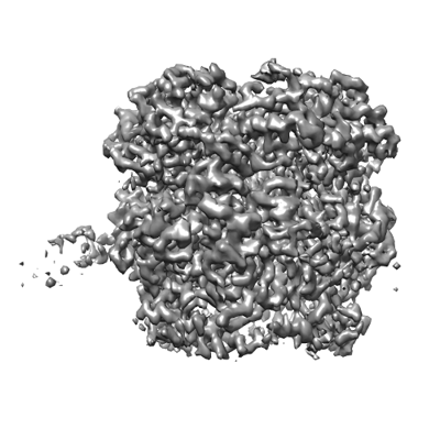
| Title | Cryo-EM structure of the proximal end of bacteriophage T5 tail, after interaction with its receptor : p142 tail terminator protein hexamer and pb6 tail tube protein trimer | |||||||||
 Map data Map data | ||||||||||
 Sample Sample |
| |||||||||
 Keywords Keywords | Bacteriophage / Siphophage / T5 / tail terminator / Trp / VIRAL PROTEIN | |||||||||
| Function / homology |  Function and homology information Function and homology informationvirus tail, tube / symbiont genome ejection through host cell envelope, long flexible tail mechanism / viral tail assembly / virus tail / viral release from host cell by cytolysis Similarity search - Function | |||||||||
| Biological species |  Escherichia phage T5 (virus) / Escherichia virus T5 Escherichia phage T5 (virus) / Escherichia virus T5 | |||||||||
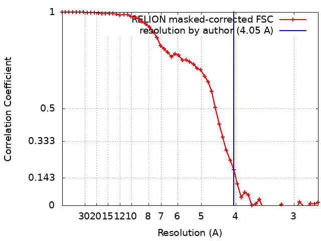
| Method | single particle reconstruction / cryo EM / Resolution: 4.05 Å | |||||||||
 Authors Authors | Linares R / Effantin G / Breyton C / Darnault C / Epalle N / Boeri Erba E / Schoehn G | |||||||||
| Funding support |  France, 2 items France, 2 items
| |||||||||
 Citation Citation | Journal: J Virol / Year: 2025 Title: About bacteriophage tail terminator and tail completion proteins: structure of the proximal extremity of siphophage T5 tail. Authors: Romain Linares / Cécile Breyton / Abstract: Bacteriophages are viruses infecting bacteria. The vast majority of them bear a tail, allowing host recognition, cell wall perforation, and DNA injection into the host cytoplasm. Using electron cryo- ...Bacteriophages are viruses infecting bacteria. The vast majority of them bear a tail, allowing host recognition, cell wall perforation, and DNA injection into the host cytoplasm. Using electron cryo-microscopy (cryo-EM) and single particle analysis, we determined the organization of the tail proximal extremity of siphophage T5 that possesses a long flexible tail and solved the structure of its tail terminator protein p142 (TrP). It allowed us to confirm the common evolutionary origin between T5 TrP and other known or putative TrPs from siphophages, myophages, and bacterial tail-like machines, despite very poor sequence conservation. By also determining the structure of the T5 tail proximal extremity after interaction with T5 bacterial receptor FhuA, we showed that no conformational changes occur in TrP and confirmed that the infection signal transduction is not carried by the tube itself. We also investigated the location of T5 Neck1 or tail completion protein p143 (TCP) and showed, thanks to a combination of cryo-EM and structure prediction using Alphafold2, that it is not located at the capsid-to-tail interface as suggested by its position in the genome, but instead, very unexpectedly, on the side of T5 tail tip, and that it appears to be monomeric. Based on structure comparison with other putative TCPs predicted structures, this feature could not be shared by other TCPs and questions the affiliation of p143 to this family of protein.IMPORTANCEBacteriophages, viruses infecting bacteria, are the most abundant living entities on Earth. They are present in all ecosystems where bacteria develop and are instrumental in the regulation, diversity, evolution, and pathogeny of microbial populations. Moreover, with the increasing number of pathogenic strains resistant to antibiotics, virulent phages are considered a serious alternative or complement to classical treatments. 96% of all phages present a tail that allows host recognition and safe channeling of the DNA to the host cytoplasm. We present the atomic model of the proximal extremity of the siphophage T5 tail, confirming structural similarities with other phages. This structure, combined with results previously published and further explored, also allowed a review and a discussion on the role and localization of a mysterious tail protein, the tail completion protein, which is known to be present in the phage tails, but that was never identified in a phage structure. | |||||||||
| History |
|
- Structure visualization
| Supplemental images |
|---|
- Downloads & links
-EMDB archive
| Map data | emd_15968.map.gz | 2 MB | EMDB map data format | |
|---|---|---|---|---|
| Header (meta data) | emd-15968-v30.xmlemd-15968.xml | 21.4 KB 21.4 KB | Display Display | EMDB header |
| FSC (resolution estimation) | emd_15968_fsc.xml | 5.1 KB | Display | FSC data file |
| Images |  emd_15968.png emd_15968.png | 101.1 KB | ||
| Masks | emd_15968_msk_1.map | 10.5 MB | Mask map | |
| Filedesc metadata | emd-15968.cif.gz | 6.8 KB | ||
| Others | emd_15968_additional_1.map.gzemd_15968_half_map_1.map.gzemd_15968_half_map_2.map.gz | 7.9 MB 7.9 MB 7.9 MB | ||
| Archive directory |  http://ftp.pdbj.org/pub/emdb/structures/EMD-15968ftp://ftp.pdbj.org/pub/emdb/structures/EMD-15968 http://ftp.pdbj.org/pub/emdb/structures/EMD-15968ftp://ftp.pdbj.org/pub/emdb/structures/EMD-15968 | HTTPS FTP |
-Related structure data
| Related structure data |  8bcuMC  8bcpC M: atomic model generated by this map C: citing same article ( |
|---|---|
| Similar structure data |
-Links
| EMDB pages | EMDB (EBI/PDBe) / EMDataResource |
|---|
-Map
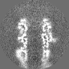
| File | Download / File: emd_15968.map.gz / Format: CCP4 / Size: 10.5 MB / Type: IMAGE STORED AS FLOATING POINT NUMBER (4 BYTES) | ||||||||||||||||||||||||||||||||||||
|---|---|---|---|---|---|---|---|---|---|---|---|---|---|---|---|---|---|---|---|---|---|---|---|---|---|---|---|---|---|---|---|---|---|---|---|---|---|
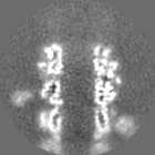
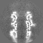
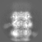
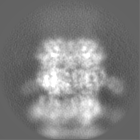
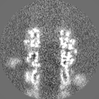
| Projections & slices | Image control
Images are generated by Spider. | ||||||||||||||||||||||||||||||||||||
| Voxel size | X=Y=Z: 1.351 Å | ||||||||||||||||||||||||||||||||||||
| Density |
| ||||||||||||||||||||||||||||||||||||
| Symmetry | Space group: 1 | ||||||||||||||||||||||||||||||||||||
| Details | EMDB XML:
|
 Z (Sec.)
 Y (Row.)
 X (Col.)


-Supplemental data
-Mask #1
| File | emd_15968_msk_1.map | ||||||||||||
|---|---|---|---|---|---|---|---|---|---|---|---|---|---|
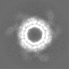
| Projections & Slices |
| ||||||||||||
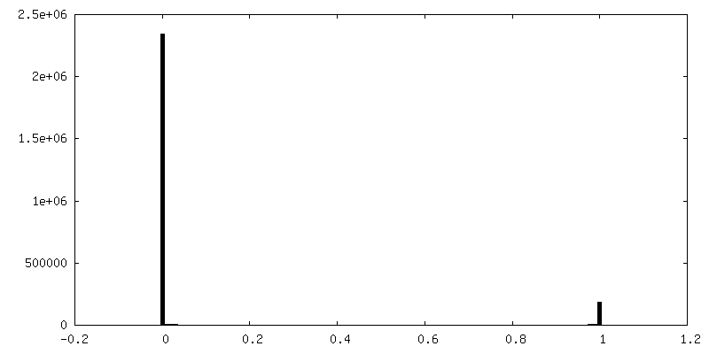
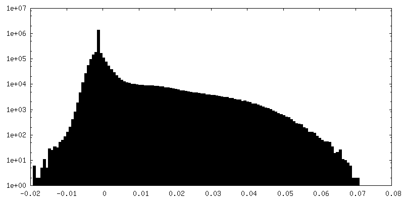
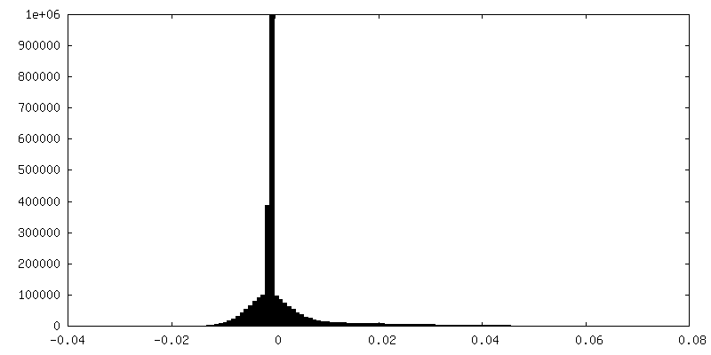
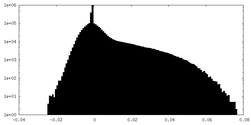
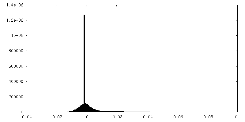
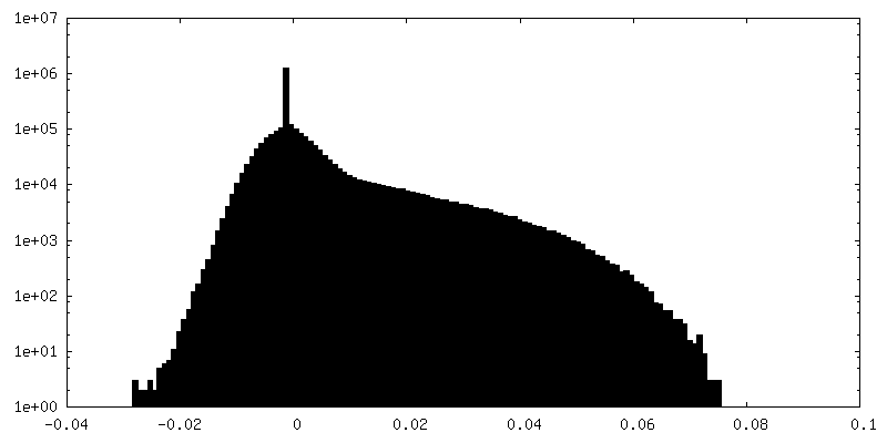
| Density Histograms |


-Additional map: #1
| File | emd_15968_additional_1.map | ||||||||||||
|---|---|---|---|---|---|---|---|---|---|---|---|---|---|
| Projections & Slices |
| ||||||||||||
| Density Histograms |


-Half map: #1
| File | emd_15968_half_map_1.map | ||||||||||||
|---|---|---|---|---|---|---|---|---|---|---|---|---|---|
| Projections & Slices |
| ||||||||||||
| Density Histograms |


-Half map: #2
| File | emd_15968_half_map_2.map | ||||||||||||
|---|---|---|---|---|---|---|---|---|---|---|---|---|---|
| Projections & Slices |
| ||||||||||||
| Density Histograms |


- Sample components
-Entire : Escherichia virus T5
| Entire | Name: Escherichia virus T5 |
|---|---|
| Components |
|
-Supramolecule #1: Escherichia virus T5
| Supramolecule | Name: Escherichia virus T5 / type: virus / ID: 1 / Parent: 0 / Macromolecule list: all Details: Pure T5 tails obtained by infecting E. coli F strain with the amber mutant phage T5D20am30d, incubated with the bacterial receptor FhuA reconstituted in nanodiscs NCBI-ID: 2695836 / Sci species name: Escherichia virus T5 / Sci species strain: T5D20am30d / Virus type: VIRUS-LIKE PARTICLE / Virus isolate: STRAIN / Virus enveloped: No / Virus empty: No |
|---|---|
| Host (natural) | Organism:  |
-Macromolecule #1: Tail tube terminator protein p142
| Macromolecule | Name: Tail tube terminator protein p142 / type: protein_or_peptide / ID: 1 / Number of copies: 6 / Enantiomer: LEVO |
|---|---|
| Source (natural) | Organism: Escherichia phage T5 (virus) |
| Molecular weight | Theoretical: 18.378643 KDa |
| Sequence | String: MDHRTSIAQA MVDRISKQMD GSQPDEYFNN LYGNVSRQTY KFEEIREFPY VAVHIGTETG QYLPSGQQWM FLELPILVYD KEKTDIQEQ LEKLVADIKT VIDTGGNLEY TVSKPNGSTF PCEATDMIIT SVSTDEGLLA PYGLAEINVT VRYQPPRRSL R R UniProtKB: Tail tube terminator protein p142 |
-Macromolecule #2: Tail tube protein
| Macromolecule | Name: Tail tube protein / type: protein_or_peptide / ID: 2 / Number of copies: 3 / Enantiomer: LEVO |
|---|---|
| Source (natural) | Organism: Escherichia phage T5 (virus) |
| Molecular weight | Theoretical: 50.459215 KDa |
| Sequence | String: MSLQLLRNTR IFVSTVKTGH NKTNTQEILV QDDISWGQDS NSTDITVNEA GPRPTRGSKR FNDSLNAAEW SFSTYILPYK DKNTSKQIV PDYMLWHALS SGRAINLEGT TGAHNNATNF MVNFKDNSYH ELAMLHIYIL TDKTWSYIDS CQINQAEVNV D IEDIGRVT ...String: MSLQLLRNTR IFVSTVKTGH NKTNTQEILV QDDISWGQDS NSTDITVNEA GPRPTRGSKR FNDSLNAAEW SFSTYILPYK DKNTSKQIV PDYMLWHALS SGRAINLEGT TGAHNNATNF MVNFKDNSYH ELAMLHIYIL TDKTWSYIDS CQINQAEVNV D IEDIGRVT WSGNGNQLIP LDEQPFDPDQ IGIDDETYMT IQGSYIKNKL TILKIKDMDT NKSYDIPITG GTFTINNNIT YL TPNVMSR VTIPIGSFTG AFELTGSLTA YLNDKSLGSM ELYKDLIKTL KVVNRFEIAL VLGGEYDDER PAAILVAKQA HVN IPTIET DDVLGTSVEF KAIPSDLDAG DEGYLGFSSK YTRTTINNLI VNGDGATDAV TAITVKSAGN VTTLNRSATL QMSV EVTPS SARNKEVTWA ITAGDAATIN ATGLLRADAS KTGAVTVEAT AKDGSGVKGT KVITVTAGG UniProtKB: Tail tube protein pb6 |
-Experimental details
-Structure determination
| Method | cryo EM |
|---|---|
 Processing Processing | single particle reconstruction |
| Aggregation state | particle |
-Sample preparation
| Buffer | pH: 8 Component:
| |||||||||||||||
|---|---|---|---|---|---|---|---|---|---|---|---|---|---|---|---|---|
| Grid | Model: Quantifoil R2/1 / Material: COPPER/RHODIUM / Mesh: 300 / Support film - Material: CARBON / Support film - topology: HOLEY ARRAY / Pretreatment - Type: GLOW DISCHARGE / Pretreatment - Time: 30 sec. / Details: 25 mA | |||||||||||||||
| Vitrification | Cryogen name: ETHANE / Chamber humidity: 100 % / Chamber temperature: 293.15 K / Instrument: FEI VITROBOT MARK IV Details: 3 uL of T5 tails + FhuA-nanodisc sample were deposited on a freshly glow discharged EM grid and plunge-frozen in nitrogen-cooled liquid ethane using a ThermoFisher Mark IV Vitrobot device ...Details: 3 uL of T5 tails + FhuA-nanodisc sample were deposited on a freshly glow discharged EM grid and plunge-frozen in nitrogen-cooled liquid ethane using a ThermoFisher Mark IV Vitrobot device (100 percent humidity, 20 Celsius degrees, 5 s blotting time, blot force 0). | |||||||||||||||
| Details | Pure T5 tails obtained by infecting E. coli F strain with the amber mutant phage T5D20am30d, incubated with the bacterial receptor FhuA reconstituted in nanodiscs |
- Electron microscopy
| Microscope | FEI TITAN KRIOS |
|---|---|
| Image recording | Film or detector model: GATAN K2 SUMMIT (4k x 4k) / Detector mode: COUNTING / Average electron dose: 40.0 e/Å2 |
| Electron beam | Acceleration voltage: 300 kV / Electron source:  FIELD EMISSION GUN FIELD EMISSION GUN |
| Electron optics | Illumination mode: FLOOD BEAM / Imaging mode: BRIGHT FIELD / Cs: 2.7 mm / Nominal defocus max: 3.0 µm / Nominal defocus min: 1.0 µm / Nominal magnification: 105000 |
| Sample stage | Specimen holder model: FEI TITAN KRIOS AUTOGRID HOLDER / Cooling holder cryogen: NITROGEN |
| Experimental equipment |  Model: Titan Krios / Image courtesy: FEI Company |
+Image processing
-Atomic model buiding 1
| Refinement | Space: REAL / Protocol: BACKBONE TRACE / Overall B value: 120 |
|---|---|
| Output model | PDB-8bcu: |
