 Movie
Movie Controller

[English] 日本語
 Yorodumi
- PDB-8bb7: Crystal structure of Mouse Plexin-B1 (20-535) in complex with VHH15 -
+ Open data

- Basic information
| Entry | Database: PDB / ID: 8bb7 | ||||||
|---|---|---|---|---|---|---|---|
| Title | Crystal structure of Mouse Plexin-B1 (20-535) in complex with VHH15 | ||||||
 Components Components |
| ||||||
 Keywords Keywords | SIGNALING PROTEIN / Complex / inhibitor / VHH / nanobody / Plexin / semaphorin / allosteric | ||||||
| Function / homology |  Function and homology information Function and homology informationSema4D mediated inhibition of cell attachment and migration / Sema4D induced cell migration and growth-cone collapse / : / G alpha (12/13) signalling events / negative regulation of osteoblast proliferation / semaphorin receptor complex / semaphorin receptor activity / negative regulation of cell adhesion / positive regulation of axonogenesis / inhibitory synapse assembly ...Sema4D mediated inhibition of cell attachment and migration / Sema4D induced cell migration and growth-cone collapse / : / G alpha (12/13) signalling events / negative regulation of osteoblast proliferation / semaphorin receptor complex / semaphorin receptor activity / negative regulation of cell adhesion / positive regulation of axonogenesis / inhibitory synapse assembly / GTPase activating protein binding / ossification involved in bone maturation / regulation of cytoskeleton organization / positive regulation of Rho protein signal transduction / semaphorin-plexin signaling pathway / positive regulation of GTPase activity / neuron projection morphogenesis / regulation of cell shape / postsynaptic membrane / positive regulation of phosphatidylinositol 3-kinase/protein kinase B signal transduction / glutamatergic synapse / plasma membrane Similarity search - Function | ||||||
| Biological species |  Camelidae (mammal) Camelidae (mammal) | ||||||
| Method |  X-RAY DIFFRACTION / SYNCHROTRON / MOLECULAR REPLACEMENT / Resolution: 2.95 Å X-RAY DIFFRACTION / SYNCHROTRON / MOLECULAR REPLACEMENT / Resolution: 2.95 Å | ||||||
 Authors Authors | Cowan, R. / Hall, G. / Carr, M. | ||||||
| Funding support |  United Kingdom, 1items United Kingdom, 1items
| ||||||
 Citation Citation | Journal: J.Biol.Chem. / Year: 2023 Title: Nanobody inhibitors of Plexin-B1 identify allostery in plexin-semaphorin interactions and signaling. Authors: Cowan, R. / Trokter, M. / Oleksy, A. / Fedorova, M. / Sawmynaden, K. / Worzfeld, T. / Offermanns, S. / Matthews, D. / Carr, M.D. / Hall, G. | ||||||
| History |
|
- Structure visualization
| Structure viewer | Molecule: MolmilJmol/JSmol |
|---|
- Downloads & links
-Download
| PDBx/mmCIF format | 8bb7.cif.gz | 271.7 KB | Display | PDBx/mmCIF format |
|---|---|---|---|---|
| PDB format | pdb8bb7.ent.gz | 197.2 KB | Display | PDB format |
| PDBx/mmJSON format | 8bb7.json.gz | Tree view | PDBx/mmJSON format | |
| Others |  Other downloads Other downloads |
-Validation report
| Arichive directory | https://data.pdbj.org/pub/pdb/validation_reports/bb/8bb7ftp://data.pdbj.org/pub/pdb/validation_reports/bb/8bb7 | HTTPS FTP |
|---|
-Related structure data
| Related structure data | 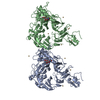 8b3kC 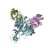 8bf4C 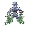 3ol2S S: Starting model for refinement C: citing same article ( |
|---|---|
| Similar structure data |
-Links
 PDBj
PDBj


- Assembly
| Deposited unit | 
| ||||||||||
|---|---|---|---|---|---|---|---|---|---|---|---|
| 1 | 
| ||||||||||
| 2 | 
| ||||||||||
| Unit cell |
|
-Components
| #1: Protein | Mass: 56450.527 Da / Num. of mol.: 2 Source method: isolated from a genetically manipulated source Source: (gene. exp.)  Homo sapiens (human) / References: UniProt: Q8CJH3 Homo sapiens (human) / References: UniProt: Q8CJH3#2: Antibody | Mass: 14258.775 Da / Num. of mol.: 2 Source method: isolated from a genetically manipulated source Details: Huarizo / Source: (gene. exp.) Camelidae (mammal) / Production host: Homo sapiens (human)#3: Sugar | ChemComp-NAG /   Type: D-saccharide, beta linking / Mass: 221.208 Da / Num. of mol.: 8 / Source method: obtained synthetically / Formula: C8H15NO6 Type: D-saccharide, beta linking / Mass: 221.208 Da / Num. of mol.: 8 / Source method: obtained synthetically / Formula: C8H15NO6#4: Water | ChemComp-HOH / |  Mass: 18.015 Da / Num. of mol.: 222 / Source method: isolated from a natural source / Formula: H2O Mass: 18.015 Da / Num. of mol.: 222 / Source method: isolated from a natural source / Formula: H2OHas ligand of interest | N | Has protein modification | Y | |
|---|
-Experimental details
-Experiment
| Experiment | Method: X-RAY DIFFRACTION / Number of used crystals: 1 |
|---|
- Sample preparation
| Crystal | Density Matthews: 3.39 Å3/Da / Density % sol: 63.8 % |
|---|---|
| Crystal grow | Temperature: 291 K / Method: vapor diffusion, hanging drop / pH: 5 Details: 8% (w/v) PEG 8000, 0.1 M Sodium Citrate, pH 5.0, 1% (w/v) dextran-sulphate (Mr 5000) |
-Data collection
| Diffraction | Mean temperature: 100 K / Serial crystal experiment: N | |||||||||||||||||||||
|---|---|---|---|---|---|---|---|---|---|---|---|---|---|---|---|---|---|---|---|---|---|---|
| Diffraction source | Source: SYNCHROTRON / Site: BESSY  / Beamline: 14.1 / Wavelength: 0.9184 Å / Beamline: 14.1 / Wavelength: 0.9184 Å | |||||||||||||||||||||
| Detector | Type: DECTRIS PILATUS 6M / Detector: PIXEL / Date: Apr 27, 2018 | |||||||||||||||||||||
| Radiation | Protocol: SINGLE WAVELENGTH / Monochromatic (M) / Laue (L): M / Scattering type: x-ray | |||||||||||||||||||||
| Radiation wavelength | Wavelength: 0.9184 Å / Relative weight: 1 | |||||||||||||||||||||
| Reflection | Resolution: 2.948→47.194 Å / Num. obs: 36092 / % possible obs: 99.9 % / Redundancy: 6.4 % / Biso Wilson estimate: 42.66 Å2 / CC1/2: 0.953 / Rmerge(I) obs: 0.42 / Rpim(I) all: 0.274 / Rrim(I) all: 0.504 / Net I/σ(I): 5.6 | |||||||||||||||||||||
| Reflection shell | Diffraction-ID: 1
|
- Processing
| Software |
| |||||||||||||||||||||||||||||||||||||||||||||||||||||||||||||||||||||||||||||||||||||||||||
|---|---|---|---|---|---|---|---|---|---|---|---|---|---|---|---|---|---|---|---|---|---|---|---|---|---|---|---|---|---|---|---|---|---|---|---|---|---|---|---|---|---|---|---|---|---|---|---|---|---|---|---|---|---|---|---|---|---|---|---|---|---|---|---|---|---|---|---|---|---|---|---|---|---|---|---|---|---|---|---|---|---|---|---|---|---|---|---|---|---|---|---|---|
| Refinement | Method to determine structure: MOLECULAR REPLACEMENT Starting model: 3OL2 Resolution: 2.95→47.15 Å / SU ML: 0.3244 / Cross valid method: FREE R-VALUE / σ(F): 1.33 / Phase error: 23.0364 Stereochemistry target values: GeoStd + Monomer Library + CDL v1.2
| |||||||||||||||||||||||||||||||||||||||||||||||||||||||||||||||||||||||||||||||||||||||||||
| Solvent computation | Shrinkage radii: 0.9 Å / VDW probe radii: 1.1 Å / Solvent model: FLAT BULK SOLVENT MODEL | |||||||||||||||||||||||||||||||||||||||||||||||||||||||||||||||||||||||||||||||||||||||||||
| Displacement parameters | Biso mean: 43.18 Å2 | |||||||||||||||||||||||||||||||||||||||||||||||||||||||||||||||||||||||||||||||||||||||||||
| Refinement step | Cycle: LAST / Resolution: 2.95→47.15 Å
| |||||||||||||||||||||||||||||||||||||||||||||||||||||||||||||||||||||||||||||||||||||||||||
| Refine LS restraints |
| |||||||||||||||||||||||||||||||||||||||||||||||||||||||||||||||||||||||||||||||||||||||||||
| LS refinement shell |
|
