 Movie
Movie Controller


+ Open data

- Basic information
| Entry | Database: PDB / ID: 8b5w | ||||||
|---|---|---|---|---|---|---|---|
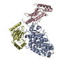
| Title | Crystal structure of the E3 module from UBR4 | ||||||
 Components Components | cDNA FLJ12511 fis, clone NT2RM2001727, highly similar to Homo sapiens ubiquitin protein ligase E3 component n-recognin 4 (UBR4), mRNA | ||||||
 Keywords Keywords | LIGASE / ubiquitin / E3 ligase / zinc finger | ||||||
| Function / homology | E3 ubiquitin ligase UBR4, C-terminal / E3 ubiquitin ligase UBR4-like / E3 ubiquitin-protein ligase UBR4 / UBR4 E3 catalytic module profile. / ligase activity / zinc ion binding / cDNA FLJ12511 fis, clone NT2RM2001727, highly similar to Homo sapiens ubiquitin protein ligase E3 component n-recognin 4 (UBR4), mRNA Function and homology information Function and homology information | ||||||
| Biological species |  Homo sapiens (human) Homo sapiens (human) | ||||||
| Method |  X-RAY DIFFRACTION / SYNCHROTRON / MAD / Resolution: 1.8 Å X-RAY DIFFRACTION / SYNCHROTRON / MAD / Resolution: 1.8 Å | ||||||
 Authors Authors | Virdee, S. / Mabbitt, P.D. / Barnsby-Greer, L. | ||||||
| Funding support |  United Kingdom, 1items United Kingdom, 1items
| ||||||
 Citation Citation | Journal: Nat.Struct.Mol.Biol. / Year: 2024 Title: UBE2A and UBE2B are recruited by an atypical E3 ligase module in UBR4. Authors: Barnsby-Greer, L. / Mabbitt, P.D. / Dery, M.A. / Squair, D.R. / Wood, N.T. / Lamoliatte, F. / Lange, S.M. / Virdee, S. | ||||||
| History |
|
- Structure visualization
| Structure viewer | Molecule: MolmilJmol/JSmol |
|---|
- Downloads & links
-Download
| PDBx/mmCIF format | 8b5w.cif.gz | 113.9 KB | Display | PDBx/mmCIF format |
|---|---|---|---|---|
| PDB format | pdb8b5w.ent.gz | 68.2 KB | Display | PDB format |
| PDBx/mmJSON format | 8b5w.json.gz | Tree view | PDBx/mmJSON format | |
| Others |  Other downloads Other downloads |
-Validation report
| Arichive directory | https://data.pdbj.org/pub/pdb/validation_reports/b5/8b5wftp://data.pdbj.org/pub/pdb/validation_reports/b5/8b5w | HTTPS FTP |
|---|
-Related structure data
-Links
 PDBj
PDBj

- Assembly
| Deposited unit | 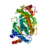
| ||||||||||||
|---|---|---|---|---|---|---|---|---|---|---|---|---|---|
| 1 |
| ||||||||||||
| Unit cell |
|
-Components
| #1: Protein | Mass: 51755.910 Da / Num. of mol.: 1 Source method: isolated from a genetically manipulated source Source: (gene. exp.) Homo sapiens (human) / Production host:  | ||||
|---|---|---|---|---|---|
| #2: Chemical | ChemComp-ZN /   Mass: 65.409 Da / Num. of mol.: 1 / Source method: obtained synthetically / Formula: Zn / Feature type: SUBJECT OF INVESTIGATION Mass: 65.409 Da / Num. of mol.: 1 / Source method: obtained synthetically / Formula: Zn / Feature type: SUBJECT OF INVESTIGATION | ||||
| #3: Chemical |   Mass: 62.068 Da / Num. of mol.: 2 / Source method: obtained synthetically / Formula: C2H6O2 Mass: 62.068 Da / Num. of mol.: 2 / Source method: obtained synthetically / Formula: C2H6O2#4: Water | ChemComp-HOH / |  Mass: 18.015 Da / Num. of mol.: 408 / Source method: isolated from a natural source / Formula: H2O Mass: 18.015 Da / Num. of mol.: 408 / Source method: isolated from a natural source / Formula: H2OHas ligand of interest | Y | |
-Experimental details
-Experiment
| Experiment | Method: X-RAY DIFFRACTION / Number of used crystals: 1 |
|---|
- Sample preparation
| Crystal | Density Matthews: 2.67 Å3/Da / Density % sol: 53.94 % |
|---|---|
| Crystal grow | Temperature: 277 K / Method: vapor diffusion, hanging drop / pH: 6.5 / Details: 10 mM Na2HPO4 13% PEG20000 |
-Data collection
| Diffraction | Mean temperature: 100 K / Serial crystal experiment: N | |||||||||
|---|---|---|---|---|---|---|---|---|---|---|
| Diffraction source | Source: SYNCHROTRON / Site: Diamond / Beamline: I24 / Wavelength: 1.2820 or 1.2831 | |||||||||
| Detector | Type: DECTRIS PILATUS3 6M / Detector: PIXEL / Date: Nov 30, 2019 | |||||||||
| Radiation | Protocol: MAD / Monochromatic (M) / Laue (L): M / Scattering type: x-ray | |||||||||
| Radiation wavelength |
| |||||||||
| Reflection | Resolution: 1.8→74.07 Å / Num. obs: 36081 / % possible obs: 91.33 % / Redundancy: 2 % / Biso Wilson estimate: 24.64 Å2 / CC1/2: 0.999 / CC star: 1 / Rmerge(I) obs: 0.02395 / Rpim(I) all: 0.02395 / Rrim(I) all: 0.03387 / Net I/σ(I): 23.84 | |||||||||
| Reflection shell | Resolution: 1.8→1.864 Å / Redundancy: 2 % / Rmerge(I) obs: 0.2907 / Mean I/σ(I) obs: 2.56 / Num. unique obs: 3577 / CC1/2: 0.872 / CC star: 0.965 / Rpim(I) all: 0.2907 / Rrim(I) all: 0.4111 / % possible all: 91.67 |
- Processing
| Software |
| |||||||||||||||||||||||||||||||||||||||||||||||||||||||||||||||||||||||||||||||||||||||||||
|---|---|---|---|---|---|---|---|---|---|---|---|---|---|---|---|---|---|---|---|---|---|---|---|---|---|---|---|---|---|---|---|---|---|---|---|---|---|---|---|---|---|---|---|---|---|---|---|---|---|---|---|---|---|---|---|---|---|---|---|---|---|---|---|---|---|---|---|---|---|---|---|---|---|---|---|---|---|---|---|---|---|---|---|---|---|---|---|---|---|---|---|---|
| Refinement | Method to determine structure: MAD / Resolution: 1.8→74.07 Å / SU ML: 0.2153 / Cross valid method: FREE R-VALUE / σ(F): 1.36 / Phase error: 24.3732 Stereochemistry target values: GeoStd + Monomer Library + CDL v1.2
| |||||||||||||||||||||||||||||||||||||||||||||||||||||||||||||||||||||||||||||||||||||||||||
| Solvent computation | Shrinkage radii: 0.9 Å / VDW probe radii: 1.11 Å / Solvent model: FLAT BULK SOLVENT MODEL | |||||||||||||||||||||||||||||||||||||||||||||||||||||||||||||||||||||||||||||||||||||||||||
| Displacement parameters | Biso mean: 28.12 Å2 | |||||||||||||||||||||||||||||||||||||||||||||||||||||||||||||||||||||||||||||||||||||||||||
| Refinement step | Cycle: LAST / Resolution: 1.8→74.07 Å
| |||||||||||||||||||||||||||||||||||||||||||||||||||||||||||||||||||||||||||||||||||||||||||
| Refine LS restraints |
| |||||||||||||||||||||||||||||||||||||||||||||||||||||||||||||||||||||||||||||||||||||||||||
| LS refinement shell |
|
