 Movie
Movie Controller

[English] 日本語
 Yorodumi
- PDB-8b53: Structure of porcine pancreatic elastase bound to a fragment of a... -
+ Open data

- Basic information
| Entry | Database: PDB / ID: 8b53 | ||||||
|---|---|---|---|---|---|---|---|
| Title | Structure of porcine pancreatic elastase bound to a fragment of a 4-azaindole inhibitor | ||||||
 Components Components | Chymotrypsin-like elastase family member 1 | ||||||
 Keywords Keywords | HYDROLASE / elastase / serine protease | ||||||
| Function / homology |  Function and homology information Function and homology informationpancreatic elastase / serine-type endopeptidase activity / proteolysis / : / metal ion binding Similarity search - Function | ||||||
| Biological species |  | ||||||
| Method |  X-RAY DIFFRACTION / SYNCHROTRON / FOURIER SYNTHESIS / Resolution: 1.25 Å X-RAY DIFFRACTION / SYNCHROTRON / FOURIER SYNTHESIS / Resolution: 1.25 Å | ||||||
 Authors Authors | Ferraroni, M. / Gerace, A. | ||||||
| Funding support | 1items
| ||||||
 Citation Citation | Journal: J.Mol.Struct. / Year: 2023 Title: X-ray structural study of human neutrophil elastase inhibition with a series of azaindoles, azaindazoles and isoxazolones Authors: Gerace, A. / Masini, V. / Crocetti, L. / Giovannoni, M.P. / Ferraroni, M. | ||||||
| History |
|
- Structure visualization
| Structure viewer | Molecule: MolmilJmol/JSmol |
|---|
- Downloads & links
-Download
| PDBx/mmCIF format | 8b53.cif.gz | 123.5 KB | Display | PDBx/mmCIF format |
|---|---|---|---|---|
| PDB format | pdb8b53.ent.gz | 92.2 KB | Display | PDB format |
| PDBx/mmJSON format | 8b53.json.gz | Tree view | PDBx/mmJSON format | |
| Others |  Other downloads Other downloads |
-Validation report
| Arichive directory | https://data.pdbj.org/pub/pdb/validation_reports/b5/8b53ftp://data.pdbj.org/pub/pdb/validation_reports/b5/8b53 | HTTPS FTP |
|---|
-Related structure data
| Related structure data | 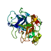 8b04C 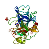 8b1yC 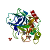 8b49C 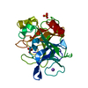 1qnjS S: Starting model for refinement C: citing same article ( |
|---|---|
| Similar structure data |
-Links
 PDBj
PDBj
- Assembly
| Deposited unit | 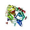
| ||||||||
|---|---|---|---|---|---|---|---|---|---|
| 1 |
| ||||||||
| Unit cell |
|
-Components
-Protein , 1 types, 1 molecules A
| #1: Protein | Mass: 28845.398 Da / Num. of mol.: 1 / Source method: isolated from a natural source / Source: (natural) |
|---|
-Non-polymers , 5 types, 333 molecules 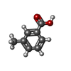


| #2: Chemical | ChemComp-OVV /  Mass: 136.148 Da / Num. of mol.: 1 / Source method: obtained synthetically / Formula: C8H8O2 / Feature type: SUBJECT OF INVESTIGATION Mass: 136.148 Da / Num. of mol.: 1 / Source method: obtained synthetically / Formula: C8H8O2 / Feature type: SUBJECT OF INVESTIGATION |
|---|---|
| #3: Chemical | ChemComp-SO4 /  Mass: 96.063 Da / Num. of mol.: 1 / Source method: obtained synthetically / Formula: SO4 Mass: 96.063 Da / Num. of mol.: 1 / Source method: obtained synthetically / Formula: SO4 |
| #4: Chemical | ChemComp-GOL /  Mass: 92.094 Da / Num. of mol.: 1 / Source method: obtained synthetically / Formula: C3H8O3 Mass: 92.094 Da / Num. of mol.: 1 / Source method: obtained synthetically / Formula: C3H8O3 |
| #5: Chemical | ChemComp-CA /  Mass: 40.078 Da / Num. of mol.: 1 / Source method: obtained synthetically / Formula: Ca Mass: 40.078 Da / Num. of mol.: 1 / Source method: obtained synthetically / Formula: Ca |
| #6: Water | ChemComp-HOH / Mass: 18.015 Da / Num. of mol.: 329 / Source method: isolated from a natural source / Formula: H2O |
-Details
| Has ligand of interest | Y |
|---|---|
| Has protein modification | Y |
-Experimental details
-Experiment
| Experiment | Method: X-RAY DIFFRACTION / Number of used crystals: 1 |
|---|
- Sample preparation
| Crystal | Density Matthews: 1.81 Å3/Da / Density % sol: 31.99 % |
|---|---|
| Crystal grow | Temperature: 296 K / Method: vapor diffusion, hanging drop / pH: 5.1 / Details: sodium sulfate, sodium acetate |
-Data collection
| Diffraction | Mean temperature: 100 K / Serial crystal experiment: N | ||||||||||||||||||||||||||||||
|---|---|---|---|---|---|---|---|---|---|---|---|---|---|---|---|---|---|---|---|---|---|---|---|---|---|---|---|---|---|---|---|
| Diffraction source | Source: SYNCHROTRON / Site: ELETTRA  / Beamline: 11.2C / Wavelength: 0.9999 Å / Beamline: 11.2C / Wavelength: 0.9999 Å | ||||||||||||||||||||||||||||||
| Detector | Type: DECTRIS PILATUS 6M / Detector: PIXEL / Date: Oct 8, 2020 | ||||||||||||||||||||||||||||||
| Radiation | Protocol: SINGLE WAVELENGTH / Monochromatic (M) / Laue (L): M / Scattering type: x-ray | ||||||||||||||||||||||||||||||
| Radiation wavelength | Wavelength: 0.9999 Å / Relative weight: 1 | ||||||||||||||||||||||||||||||
| Reflection | Resolution: 1.25→22.25 Å / Num. obs: 60617 / % possible obs: 100 % / Redundancy: 11.3 % / CC1/2: 0.997 / Rmerge(I) obs: 0.103 / Rpim(I) all: 0.032 / Rrim(I) all: 0.108 / Net I/σ(I): 11.3 / Num. measured all: 685669 / Scaling rejects: 20516 | ||||||||||||||||||||||||||||||
| Reflection shell | Diffraction-ID: 1
|
- Processing
| Software |
| |||||||||||||||||||||||||||||||||||||||||||||||||||||||||||||||||
|---|---|---|---|---|---|---|---|---|---|---|---|---|---|---|---|---|---|---|---|---|---|---|---|---|---|---|---|---|---|---|---|---|---|---|---|---|---|---|---|---|---|---|---|---|---|---|---|---|---|---|---|---|---|---|---|---|---|---|---|---|---|---|---|---|---|---|
| Refinement | Method to determine structure: FOURIER SYNTHESIS Starting model: 1QNJ Resolution: 1.25→22.25 Å / Cor.coef. Fo:Fc: 0.981 / Cor.coef. Fo:Fc free: 0.974 / SU B: 0.975 / SU ML: 0.02 / SU R Cruickshank DPI: 0.0345 / Cross valid method: THROUGHOUT / σ(F): 0 / ESU R: 0.034 / ESU R Free: 0.034 / Stereochemistry target values: MAXIMUM LIKELIHOOD Details: HYDROGENS HAVE BEEN ADDED IN THE RIDING POSITIONS U VALUES : REFINED INDIVIDUALLY
| |||||||||||||||||||||||||||||||||||||||||||||||||||||||||||||||||
| Solvent computation | Ion probe radii: 0.8 Å / Shrinkage radii: 0.8 Å / VDW probe radii: 1.2 Å / Solvent model: MASK | |||||||||||||||||||||||||||||||||||||||||||||||||||||||||||||||||
| Displacement parameters | Biso max: 81.82 Å2 / Biso mean: 11.145 Å2 / Biso min: 3.2 Å2
| |||||||||||||||||||||||||||||||||||||||||||||||||||||||||||||||||
| Refinement step | Cycle: final / Resolution: 1.25→22.25 Å
| |||||||||||||||||||||||||||||||||||||||||||||||||||||||||||||||||
| Refine LS restraints |
| |||||||||||||||||||||||||||||||||||||||||||||||||||||||||||||||||
| LS refinement shell | Resolution: 1.25→1.282 Å / Rfactor Rfree error: 0 / Total num. of bins used: 20
|
