Peptidase M73, camelysin / Camelysin metallo-endopeptidase / Signal peptide, camelysin / sporulation resulting in formation of a cellular spore / extracellular region / identical protein binding / Major biofilm matrix component
Function and homology information
Biological species
Bacillus subtilis (bacteria)
Method
ELECTRON MICROSCOPY / helical reconstruction / cryo EM / Resolution: 3.47 Å
Journal: Nat Commun / Year: 2022 Title: Donor-strand exchange drives assembly of the TasA scaffold in Bacillus subtilis biofilms. Authors: Jan Böhning / Mnar Ghrayeb / Conrado Pedebos / Daniel K Abbas / Syma Khalid / Liraz Chai / Tanmay A M Bharat / Abstract: Many bacteria in nature exist in multicellular communities termed biofilms, where cells are embedded in an extracellular matrix that provides rigidity to the biofilm and protects cells from chemical ...Many bacteria in nature exist in multicellular communities termed biofilms, where cells are embedded in an extracellular matrix that provides rigidity to the biofilm and protects cells from chemical and mechanical stresses. In the Gram-positive model bacterium Bacillus subtilis, TasA is the major protein component of the biofilm matrix, where it has been reported to form functional amyloid fibres contributing to biofilm structure and stability. Here, we present electron cryomicroscopy structures of TasA fibres, which show that, rather than forming amyloid fibrils, TasA monomers assemble into fibres through donor-strand exchange, with each subunit donating a β-strand to complete the fold of the next subunit along the fibre. Combining electron cryotomography, atomic force microscopy, and mutational studies, we show how TasA fibres congregate in three dimensions to form abundant fibre bundles that are essential for B. subtilis biofilm formation. Our study explains the previously observed biochemical properties of TasA and shows how a bacterial extracellular globular protein can assemble from monomers into β-sheet-rich fibres, and how such fibres assemble into bundles in biofilms.
Data content type: EM metadata / Data content type: EM metadata / EM metadata / Group: Data processing / Experimental summary / Data content type: EM metadata / EM metadata / Category: em_admin / em_software / Data content type: EM metadata / EM metadata / Item: _em_admin.last_update / _em_software.name
In the structure databanks used in Yorodumi, some data are registered as the other names, "COVID-19 virus" and "2019-nCoV". Here are the details of the virus and the list of structure data.
Jan 31, 2019. EMDB accession codes are about to change! (news from PDBe EMDB page)
EMDB accession codes are about to change! (news from PDBe EMDB page)
The allocation of 4 digits for EMDB accession codes will soon come to an end. Whilst these codes will remain in use, new EMDB accession codes will include an additional digit and will expand incrementally as the available range of codes is exhausted. The current 4-digit format prefixed with “EMD-” (i.e. EMD-XXXX) will advance to a 5-digit format (i.e. EMD-XXXXX), and so on. It is currently estimated that the 4-digit codes will be depleted around Spring 2019, at which point the 5-digit format will come into force.
The EM Navigator/Yorodumi systems omit the EMD- prefix.
Related info.:Q: What is EMD? / ID/Accession-code notation in Yorodumi/EM Navigator
Yorodumi is a browser for structure data from EMDB, PDB, SASBDB, etc.
This page is also the successor to EM Navigator detail page, and also detail information page/front-end page for Omokage search.
The word "yorodu" (or yorozu) is an old Japanese word meaning "ten thousand". "mi" (miru) is to see.
Related info.:EMDB / PDB / SASBDB / Comparison of 3 databanks / Yorodumi Search / Aug 31, 2016. New EM Navigator & Yorodumi / Yorodumi Papers / Jmol/JSmol / Function and homology information / Changes in new EM Navigator and Yorodumi
 Movie
Movie Controller


 Open data
 Basic information
 Components
 Keywords
 Function and homology information
 Authors
 United Kingdom, 1items
  Citation
 Structure visualization
 Downloads & links
 Other downloads
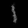
 PDBj
PDBj Assembly
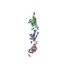
 Sample preparation
 Electron microscopy imaging
 FIELD EMISSION GUN / Accelerating voltage: 300 kV / Illumination mode: FLOOD BEAM
 Processing