 Movie
Movie Controller


+ Open data

- Basic information
| Entry | 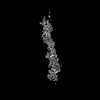 | |||||||||
|---|---|---|---|---|---|---|---|---|---|---|
| Title | Cryo-EM structure of a TasA fibre | |||||||||
 Map data Map data | Helical reconstruction cryo-EM map of TasA fibres. | |||||||||
 Sample Sample |
| |||||||||
 Keywords Keywords | Biofilm matrix fibre / PROTEIN FIBRIL | |||||||||
| Function / homology | Peptidase M73, camelysin / Camelysin metallo-endopeptidase / Signal peptide, camelysin / sporulation resulting in formation of a cellular spore / extracellular region / identical protein binding / Major biofilm matrix component Function and homology information Function and homology information | |||||||||
| Biological species |  | |||||||||
| Method | helical reconstruction / cryo EM / Resolution: 3.47 Å | |||||||||
 Authors Authors | Boehning J / Bharat TAM | |||||||||
| Funding support |  United Kingdom, 1 items United Kingdom, 1 items
| |||||||||
 Citation Citation | Journal: Nat Commun / Year: 2022 Title: Donor-strand exchange drives assembly of the TasA scaffold in Bacillus subtilis biofilms. Authors: Jan Böhning / Mnar Ghrayeb / Conrado Pedebos / Daniel K Abbas / Syma Khalid / Liraz Chai / Tanmay A M Bharat /  Abstract: Many bacteria in nature exist in multicellular communities termed biofilms, where cells are embedded in an extracellular matrix that provides rigidity to the biofilm and protects cells from chemical ...Many bacteria in nature exist in multicellular communities termed biofilms, where cells are embedded in an extracellular matrix that provides rigidity to the biofilm and protects cells from chemical and mechanical stresses. In the Gram-positive model bacterium Bacillus subtilis, TasA is the major protein component of the biofilm matrix, where it has been reported to form functional amyloid fibres contributing to biofilm structure and stability. Here, we present electron cryomicroscopy structures of TasA fibres, which show that, rather than forming amyloid fibrils, TasA monomers assemble into fibres through donor-strand exchange, with each subunit donating a β-strand to complete the fold of the next subunit along the fibre. Combining electron cryotomography, atomic force microscopy, and mutational studies, we show how TasA fibres congregate in three dimensions to form abundant fibre bundles that are essential for B. subtilis biofilm formation. Our study explains the previously observed biochemical properties of TasA and shows how a bacterial extracellular globular protein can assemble from monomers into β-sheet-rich fibres, and how such fibres assemble into bundles in biofilms. #1: Journal: Biorxiv / Year: 2022Title: Molecular architecture of the TasA biofilm scaffold in Bacillus subtilis Authors: Bohning J / Ghrayeb M / Pedebos C / Abbas DK / Khalid S / Chai L / Bharat TAM | |||||||||
| History |
|
- Structure visualization
| Supplemental images |
|---|

- Downloads & links
-EMDB archive
| Map data | emd_15673.map.gz | 5 MB | EMDB map data format | |
|---|---|---|---|---|
| Header (meta data) | emd-15673-v30.xmlemd-15673.xml | 18.9 KB 18.9 KB | Display Display | EMDB header |
| FSC (resolution estimation) | emd_15673_fsc.xml | 10 KB | Display | FSC data file |
| Images |  emd_15673.png emd_15673.png | 65.8 KB | ||
| Masks | emd_15673_msk_1.map | 83.7 MB | Mask map | |
| Filedesc metadata | emd-15673.cif.gz | 5.9 KB | ||
| Others | emd_15673_half_map_1.map.gzemd_15673_half_map_2.map.gz | 65.4 MB 65.4 MB | ||
| Archive directory |  http://ftp.pdbj.org/pub/emdb/structures/EMD-15673ftp://ftp.pdbj.org/pub/emdb/structures/EMD-15673 http://ftp.pdbj.org/pub/emdb/structures/EMD-15673ftp://ftp.pdbj.org/pub/emdb/structures/EMD-15673 | HTTPS FTP |
-Related structure data
| Related structure data | 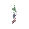 8aurMC M: atomic model generated by this map C: citing same article ( |
|---|---|
| Similar structure data |
-Links
| EMDB pages | EMDB (EBI/PDBe) / EMDataResource |
|---|
-Map
| File | Download / File: emd_15673.map.gz / Format: CCP4 / Size: 83.7 MB / Type: IMAGE STORED AS FLOATING POINT NUMBER (4 BYTES) | ||||||||||||||||||||||||||||||||||||
|---|---|---|---|---|---|---|---|---|---|---|---|---|---|---|---|---|---|---|---|---|---|---|---|---|---|---|---|---|---|---|---|---|---|---|---|---|---|
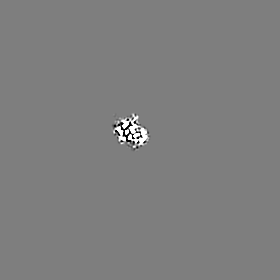
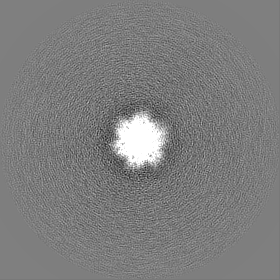
| Annotation | Helical reconstruction cryo-EM map of TasA fibres. | ||||||||||||||||||||||||||||||||||||
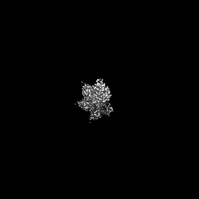
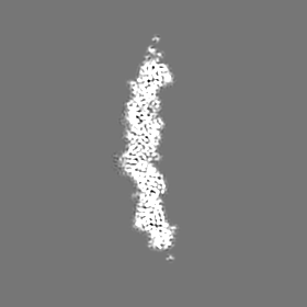
| Projections & slices | Image control
Images are generated by Spider. | ||||||||||||||||||||||||||||||||||||
| Voxel size | X=Y=Z: 1.092 Å | ||||||||||||||||||||||||||||||||||||
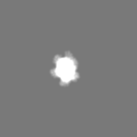
| Density |
| ||||||||||||||||||||||||||||||||||||
| Symmetry | Space group: 1 | ||||||||||||||||||||||||||||||||||||
| Details | EMDB XML:
|
 Z (Sec.)
 Y (Row.)
 X (Col.)


-Supplemental data
-Mask #1
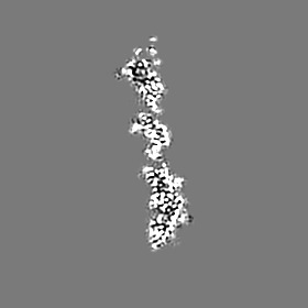
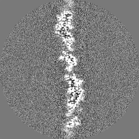
| File | emd_15673_msk_1.map | ||||||||||||
|---|---|---|---|---|---|---|---|---|---|---|---|---|---|
| Projections & Slices |
| ||||||||||||
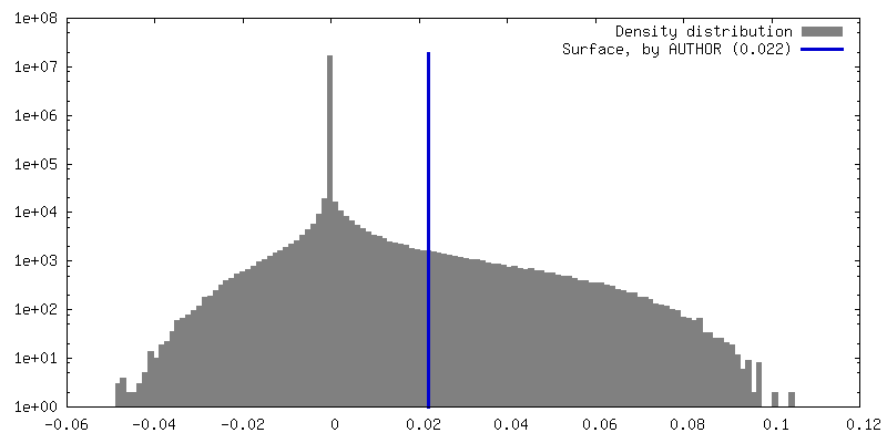
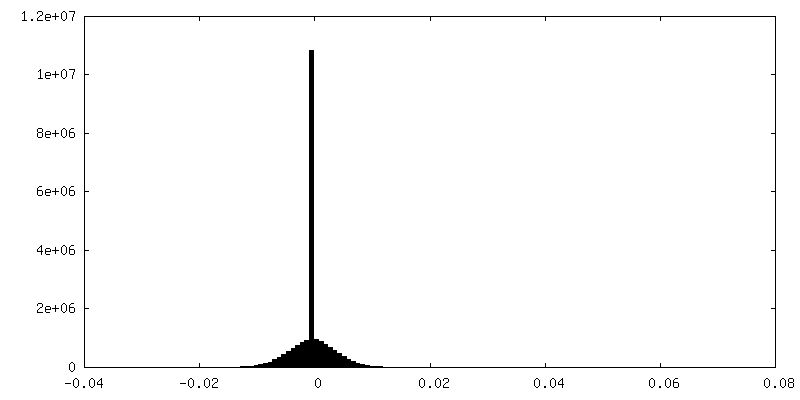
| Density Histograms |


-Half map: #2
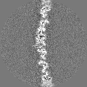
| File | emd_15673_half_map_1.map | ||||||||||||
|---|---|---|---|---|---|---|---|---|---|---|---|---|---|
| Projections & Slices |
| ||||||||||||
| Density Histograms |


-Half map: #1
| File | emd_15673_half_map_2.map | ||||||||||||
|---|---|---|---|---|---|---|---|---|---|---|---|---|---|
| Projections & Slices |
| ||||||||||||
| Density Histograms |


- Sample components
-Entire : Three-subunit TasA filament
| Entire | Name: Three-subunit TasA filament |
|---|---|
| Components |
|
-Supramolecule #1: Three-subunit TasA filament
| Supramolecule | Name: Three-subunit TasA filament / type: complex / ID: 1 / Parent: 0 / Macromolecule list: all |
|---|---|
| Source (natural) | Organism: |
| Molecular weight | Theoretical: 77.211 KDa |
-Macromolecule #1: Major biofilm matrix component
| Macromolecule | Name: Major biofilm matrix component / type: protein_or_peptide / ID: 1 / Number of copies: 3 / Enantiomer: LEVO |
|---|---|
| Source (natural) | Organism: |
| Molecular weight | Theoretical: 25.767654 KDa |
| Sequence | String: AFNDIKSKDA TFASGTLDLS AKENSASVNL SNLKPGDKLT KDFQFENNGS LAIKEVLMAL NYGDFKANGG SNTSPEDFLS QFEVTLLTV GKEGGNGYPK NIILDDANLK DLYLMSAKND AAAAEKIKKQ IDPKFLNASG KVNVATIDGK TAPEYDGVPK T PTDFDQVQ ...String: AFNDIKSKDA TFASGTLDLS AKENSASVNL SNLKPGDKLT KDFQFENNGS LAIKEVLMAL NYGDFKANGG SNTSPEDFLS QFEVTLLTV GKEGGNGYPK NIILDDANLK DLYLMSAKND AAAAEKIKKQ IDPKFLNASG KVNVATIDGK TAPEYDGVPK T PTDFDQVQ MEIQFKDDKT KDEKGLMVQN KYQGNSIKLQ FSFEATQWNG LTIKKDHTDK DGYVKENEKA HSEDKN UniProtKB: Major biofilm matrix component |
-Experimental details
-Structure determination
| Method | cryo EM |
|---|---|
 Processing Processing | helical reconstruction |
| Aggregation state | filament |
-Sample preparation
| Buffer | pH: 8 |
|---|---|
| Grid | Model: Quantifoil R2/2 / Material: COPPER/RHODIUM / Mesh: 200 / Support film - Material: CARBON / Support film - topology: HOLEY ARRAY / Pretreatment - Type: GLOW DISCHARGE / Pretreatment - Time: 20 sec. |
| Vitrification | Cryogen name: ETHANE |
- Electron microscopy
| Microscope | FEI TITAN KRIOS |
|---|---|
| Image recording | Film or detector model: GATAN K3 BIOQUANTUM (6k x 4k) / Average electron dose: 48.5 e/Å2 |
| Electron beam | Acceleration voltage: 300 kV / Electron source:  FIELD EMISSION GUN FIELD EMISSION GUN |
| Electron optics | Illumination mode: FLOOD BEAM / Imaging mode: BRIGHT FIELD / Cs: 2.7 mm / Nominal defocus max: 2.0 µm / Nominal defocus min: 1.0 µm |
| Sample stage | Specimen holder model: FEI TITAN KRIOS AUTOGRID HOLDER / Cooling holder cryogen: NITROGEN |
| Experimental equipment |  Model: Titan Krios / Image courtesy: FEI Company |
+Image processing

-Atomic model buiding 1
| Refinement | Space: REAL / Protocol: AB INITIO MODEL |
|---|---|
| Output model | PDB-8aur: |
