 Movie
Movie Controller

[English] 日本語
 Yorodumi
- PDB-8aq2: In meso structure of the membrane integral lipoprotein N-acyltran... -
+ Open data

- Basic information
| Entry | Database: PDB / ID: 8aq2 | |||||||||
|---|---|---|---|---|---|---|---|---|---|---|
| Title | In meso structure of the membrane integral lipoprotein N-acyltransferase Lnt from P. aeruginosa covalently linked with TITC | |||||||||
 Components Components | Apolipoprotein N-acyltransferase | |||||||||
 Keywords Keywords | TRANSFERASE / Lnt / apolipoprotein N-acyltransferase / Bacterial lipoproteins. | |||||||||
| Function / homology |  Function and homology information Function and homology informationapolipoprotein N-acyltransferase / : / lipoprotein biosynthetic process / plasma membrane Similarity search - Function | |||||||||
| Biological species |  Pseudomonas aeruginosa PAO1 (bacteria) Pseudomonas aeruginosa PAO1 (bacteria) | |||||||||
| Method |  X-RAY DIFFRACTION / SYNCHROTRON / MOLECULAR REPLACEMENT / Resolution: 2.6 Å X-RAY DIFFRACTION / SYNCHROTRON / MOLECULAR REPLACEMENT / Resolution: 2.6 Å | |||||||||
 Authors Authors | Huang, C.-Y. / Weichert, D. / Boland, C. / Smithers, L. / Olieric, V. / Wang, M. / Caffrey, M. | |||||||||
| Funding support |  Ireland, 2items Ireland, 2items
| |||||||||
 Citation Citation | Journal: Sci Adv / Year: 2023 Title: Structure snapshots reveal the mechanism of a bacterial membrane lipoprotein -acyltransferase. Authors: Luke Smithers / Oksana Degtjarik / Dietmar Weichert / Chia-Ying Huang / Coilín Boland / Katherine Bowen / Abraham Oluwole / Corinne Lutomski / Carol V Robinson / Eoin M Scanlan / Meitian ...Authors: Luke Smithers / Oksana Degtjarik / Dietmar Weichert / Chia-Ying Huang / Coilín Boland / Katherine Bowen / Abraham Oluwole / Corinne Lutomski / Carol V Robinson / Eoin M Scanlan / Meitian Wang / Vincent Olieric / Moran Shalev-Benami / Martin Caffrey /    Abstract: Bacterial lipoproteins (BLPs) decorate the surface of membranes in the cell envelope. They function in membrane assembly and stability, as enzymes, and in transport. The final enzyme in the BLP ...Bacterial lipoproteins (BLPs) decorate the surface of membranes in the cell envelope. They function in membrane assembly and stability, as enzymes, and in transport. The final enzyme in the BLP synthesis pathway is the apolipoprotein -acyltransferase, Lnt, which is proposed to act by a ping-pong mechanism. Here, we use x-ray crystallography and cryo-electron microscopy to chart the structural changes undergone during the progress of the enzyme through the reaction. We identify a single active site that has evolved to bind, individually and sequentially, substrates that satisfy structural and chemical criteria to position reactive parts next to the catalytic triad for reaction. This study validates the ping-pong mechanism, explains the molecular bases for Lnt's substrate promiscuity, and should facilitate the design of antibiotics with minimal off-target effects. | |||||||||
| History |
|
- Structure visualization
| Structure viewer | Molecule: MolmilJmol/JSmol |
|---|
- Downloads & links
-Download
| PDBx/mmCIF format | 8aq2.cif.gz | 235.5 KB | Display | PDBx/mmCIF format |
|---|---|---|---|---|
| PDB format | pdb8aq2.ent.gz | 187.5 KB | Display | PDB format |
| PDBx/mmJSON format | 8aq2.json.gz | Tree view | PDBx/mmJSON format | |
| Others |  Other downloads Other downloads |
-Validation report
| Arichive directory | https://data.pdbj.org/pub/pdb/validation_reports/aq/8aq2ftp://data.pdbj.org/pub/pdb/validation_reports/aq/8aq2 | HTTPS FTP |
|---|
-Related structure data
| Related structure data |  8aq3C  8aq4C  8b0kC  8b0lC  8b0mC  8b0nC  8b0oC  8b0pC  5n6mS S: Starting model for refinement C: citing same article ( |
|---|---|
| Similar structure data |


-Links
 PDBj
PDBj


- Assembly
| Deposited unit | 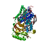
| ||||||||
|---|---|---|---|---|---|---|---|---|---|
| 1 |
| ||||||||
| Unit cell |
|
-Components
-Protein , 1 types, 1 molecules A
| #1: Protein | Mass: 58230.461 Da / Num. of mol.: 1 Source method: isolated from a genetically manipulated source Source: (gene. exp.) Pseudomonas aeruginosa PAO1 (bacteria)Strain: ATCC 15692 / DSM 22644 / CIP 104116 / JCM 14847 / LMG 12228 / 1C / PRS 101 / PAO1 Gene: lnt, cutE, PA3984 / Production host: References: UniProt: Q9ZI86, Transferases; Acyltransferases; Transferring groups other than aminoacyl groups |
|---|
-Non-polymers , 5 types, 50 molecules 


| #2: Chemical | ChemComp-OLC / (  Mass: 356.540 Da / Num. of mol.: 10 / Source method: obtained synthetically / Formula: C21H40O4 Mass: 356.540 Da / Num. of mol.: 10 / Source method: obtained synthetically / Formula: C21H40O4#3: Chemical |  Mass: 243.452 Da / Num. of mol.: 2 / Source method: obtained synthetically / Formula: C14H29NS / Feature type: SUBJECT OF INVESTIGATION Mass: 243.452 Da / Num. of mol.: 2 / Source method: obtained synthetically / Formula: C14H29NS / Feature type: SUBJECT OF INVESTIGATION#4: Chemical | ChemComp-FLC / |  Mass: 189.100 Da / Num. of mol.: 1 / Source method: obtained synthetically / Formula: C6H5O7 Mass: 189.100 Da / Num. of mol.: 1 / Source method: obtained synthetically / Formula: C6H5O7#5: Chemical | ChemComp-GOL /  Mass: 92.094 Da / Num. of mol.: 7 / Source method: obtained synthetically / Formula: C3H8O3 Mass: 92.094 Da / Num. of mol.: 7 / Source method: obtained synthetically / Formula: C3H8O3#6: Water | ChemComp-HOH / | Mass: 18.015 Da / Num. of mol.: 30 / Source method: isolated from a natural source / Formula: H2O |
|---|
-Details
| Has ligand of interest | Y |
|---|---|
| Has protein modification | Y |
-Experimental details
-Experiment
| Experiment | Method: X-RAY DIFFRACTION / Number of used crystals: 1 |
|---|
- Sample preparation
| Crystal | Density Matthews: 2.85 Å3/Da / Density % sol: 56.87 % |
|---|---|
| Crystal grow | Temperature: 293 K / Method: lipidic cubic phase Details: 30 %(v/v) PEG-500 DME, 0.1 M sodium citrate pH 5.0 and 0.1 M sodium acetate |
-Data collection
| Diffraction | Mean temperature: 100 K / Serial crystal experiment: Y |
|---|---|
| Diffraction source | Source: SYNCHROTRON / Site: SLS / Beamline: X06SA / Wavelength: 1 Å |
| Detector | Type: DECTRIS EIGER X 16M / Detector: PIXEL / Date: Mar 27, 2017 |
| Radiation | Protocol: SINGLE WAVELENGTH / Monochromatic (M) / Laue (L): M / Scattering type: x-ray |
| Radiation wavelength | Wavelength: 1 Å / Relative weight: 1 |
| Reflection | Resolution: 2.6→48.526 Å / Num. obs: 21051 / % possible obs: 99.7 % / Redundancy: 14.63 % / CC1/2: 0.99 / Rrim(I) all: 0.35 / Net I/σ(I): 6.93 |
| Reflection shell | Resolution: 2.6→2.7 Å / Redundancy: 14.59 % / Mean I/σ(I) obs: 0.85 / Num. unique obs: 2200 / CC1/2: 0.35 / Rrim(I) all: 6.29 / % possible all: 99.7 |
| Serial crystallography sample delivery | Method: fixed target |
| Serial crystallography sample delivery fixed target | Description: IMISX method / Sample dehydration prevention: COC film sandwitch / Sample holding: IMISX method/COC film / Support base: standard goniometer base |
- Processing
| Software |
| ||||||||||||||||||||||||||||||||||||||||||||||||||||||
|---|---|---|---|---|---|---|---|---|---|---|---|---|---|---|---|---|---|---|---|---|---|---|---|---|---|---|---|---|---|---|---|---|---|---|---|---|---|---|---|---|---|---|---|---|---|---|---|---|---|---|---|---|---|---|---|
| Refinement | Method to determine structure: MOLECULAR REPLACEMENT Starting model: 5N6M Resolution: 2.6→48.526 Å / SU ML: 0.42 / Cross valid method: THROUGHOUT / σ(F): 1.33 / Phase error: 30.08 / Stereochemistry target values: ML
| ||||||||||||||||||||||||||||||||||||||||||||||||||||||
| Solvent computation | Shrinkage radii: 0.9 Å / VDW probe radii: 1.11 Å / Solvent model: FLAT BULK SOLVENT MODEL | ||||||||||||||||||||||||||||||||||||||||||||||||||||||
| Displacement parameters | Biso max: 191.66 Å2 / Biso mean: 73.4173 Å2 / Biso min: 30.74 Å2 | ||||||||||||||||||||||||||||||||||||||||||||||||||||||
| Refinement step | Cycle: final / Resolution: 2.6→48.526 Å
| ||||||||||||||||||||||||||||||||||||||||||||||||||||||
| LS refinement shell | Refine-ID: X-RAY DIFFRACTION / Rfactor Rfree error: 0
| ||||||||||||||||||||||||||||||||||||||||||||||||||||||
| Refinement TLS params. | Method: refined / Origin x: -10.1329 Å / Origin y: 19.4728 Å / Origin z: -16.6794 Å
| ||||||||||||||||||||||||||||||||||||||||||||||||||||||
| Refinement TLS group |
|
