 Movie
Movie Controller

[English] 日本語
 Yorodumi
- PDB-8apy: Crystal structure of the H12A variant of the KDEL receptor bound ... -
+ Open data

- Basic information
| Entry | Database: PDB / ID: 8apy | ||||||
|---|---|---|---|---|---|---|---|
| Title | Crystal structure of the H12A variant of the KDEL receptor bound to sybody | ||||||
 Components Components |
| ||||||
 Keywords Keywords | MEMBRANE PROTEIN / Trafficking receptor / synthetic binder / KDELR | ||||||
| Function / homology |  Function and homology information Function and homology informationKDEL sequence binding / ER retention sequence binding / COPI-dependent Golgi-to-ER retrograde traffic / COPI-coated vesicle membrane / protein retention in ER lumen / COPI-mediated anterograde transport / maintenance of protein localization in endoplasmic reticulum / cis-Golgi network / retrograde vesicle-mediated transport, Golgi to endoplasmic reticulum / protein transport ...KDEL sequence binding / ER retention sequence binding / COPI-dependent Golgi-to-ER retrograde traffic / COPI-coated vesicle membrane / protein retention in ER lumen / COPI-mediated anterograde transport / maintenance of protein localization in endoplasmic reticulum / cis-Golgi network / retrograde vesicle-mediated transport, Golgi to endoplasmic reticulum / protein transport / Golgi membrane / endoplasmic reticulum membrane / endoplasmic reticulum / membrane Similarity search - Function | ||||||
| Biological species |  synthetic construct (others) | ||||||
| Method |  X-RAY DIFFRACTION / SYNCHROTRON / MOLECULAR REPLACEMENT / Resolution: 2.34 Å X-RAY DIFFRACTION / SYNCHROTRON / MOLECULAR REPLACEMENT / Resolution: 2.34 Å | ||||||
 Authors Authors | Parker, J.L. / Smith, K. / Newstead, S. | ||||||
| Funding support |  United Kingdom, 1items United Kingdom, 1items
| ||||||
 Citation Citation | Journal: Structure / Year: 2024 Title: Molecular basis for pH sensing in the KDEL trafficking receptor. Authors: Wu, Z. / Smith, K. / Gerondopoulos, A. / Sobajima, T. / Parker, J.L. / Barr, F.A. / Newstead, S. / Biggin, P.C. | ||||||
| History |
|
- Structure visualization
| Structure viewer | Molecule: MolmilJmol/JSmol |
|---|
- Downloads & links
-Download
| PDBx/mmCIF format | 8apy.cif.gz | 98.9 KB | Display | PDBx/mmCIF format |
|---|---|---|---|---|
| PDB format | pdb8apy.ent.gz | 60.2 KB | Display | PDB format |
| PDBx/mmJSON format | 8apy.json.gz | Tree view | PDBx/mmJSON format | |
| Others |  Other downloads Other downloads |
-Validation report
| Arichive directory | https://data.pdbj.org/pub/pdb/validation_reports/ap/8apyftp://data.pdbj.org/pub/pdb/validation_reports/ap/8apy | HTTPS FTP |
|---|
-Related structure data
| Related structure data | 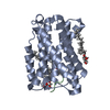 7oxeC  7oyeC  6i6jS S: Starting model for refinement C: citing same article ( |
|---|---|
| Similar structure data |
-Links
 PDBj
PDBj


- Assembly
| Deposited unit | 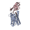
| ||||||||||||
|---|---|---|---|---|---|---|---|---|---|---|---|---|---|
| 1 |
| ||||||||||||
| Unit cell |
|
-Components
| #1: Protein | Mass: 24409.818 Da / Num. of mol.: 1 Source method: isolated from a genetically manipulated source Source: (gene. exp.)  |
|---|---|
| #2: Antibody | Mass: 13611.107 Da / Num. of mol.: 1 Source method: isolated from a genetically manipulated source Source: (gene. exp.) synthetic construct (others) / Production host:  |
| #3: Chemical | ChemComp-OLC / (  Mass: 356.540 Da / Num. of mol.: 1 / Source method: obtained synthetically / Formula: C21H40O4 Mass: 356.540 Da / Num. of mol.: 1 / Source method: obtained synthetically / Formula: C21H40O4 |
| #4: Water | ChemComp-HOH /  Mass: 18.015 Da / Num. of mol.: 17 / Source method: isolated from a natural source / Formula: H2O Mass: 18.015 Da / Num. of mol.: 17 / Source method: isolated from a natural source / Formula: H2O |
| Has ligand of interest | N |
| Has protein modification | Y |
-Experimental details
-Experiment
| Experiment | Method: X-RAY DIFFRACTION / Number of used crystals: 1 |
|---|
- Sample preparation
| Crystal | Density Matthews: 2.87 Å3/Da / Density % sol: 57.15 % |
|---|---|
| Crystal grow | Temperature: 293 K / Method: lipidic cubic phase / Details: 30% v/v PEG 400 0.1 M Tris pH 8.0. |
-Data collection
| Diffraction | Mean temperature: 100 K / Serial crystal experiment: N |
|---|---|
| Diffraction source | Source: SYNCHROTRON / Site: Diamond / Beamline: I24 / Wavelength: 0.9686 Å |
| Detector | Type: DECTRIS PILATUS 6M / Detector: PIXEL / Date: Jun 22, 2021 |
| Radiation | Protocol: SINGLE WAVELENGTH / Monochromatic (M) / Laue (L): M / Scattering type: x-ray |
| Radiation wavelength | Wavelength: 0.9686 Å / Relative weight: 1 |
| Reflection | Resolution: 2.34→42.79 Å / Num. obs: 18726 / % possible obs: 99.6 % / Redundancy: 6.2 % / Biso Wilson estimate: 56 Å2 / CC1/2: 0.997 / Rmerge(I) obs: 0.144 / Rpim(I) all: 0.094 / Net I/σ(I): 9.4 |
| Reflection shell | Resolution: 2.34→2.4 Å / Rmerge(I) obs: 1.9 / Mean I/σ(I) obs: 1 / Num. unique obs: 1371 / CC1/2: 0.406 / Rpim(I) all: 1.218 / % possible all: 99.9 |
- Processing
| Software |
| ||||||||||||||||||||||||||||||||||||||||||||||||||||||||
|---|---|---|---|---|---|---|---|---|---|---|---|---|---|---|---|---|---|---|---|---|---|---|---|---|---|---|---|---|---|---|---|---|---|---|---|---|---|---|---|---|---|---|---|---|---|---|---|---|---|---|---|---|---|---|---|---|---|
| Refinement | Method to determine structure: MOLECULAR REPLACEMENT Starting model: 6i6j Resolution: 2.34→42.79 Å / SU ML: 0.4856 / Cross valid method: FREE R-VALUE / σ(F): 1.34 / Phase error: 39.9822 Stereochemistry target values: GeoStd + Monomer Library + CDL v1.2
| ||||||||||||||||||||||||||||||||||||||||||||||||||||||||
| Solvent computation | Shrinkage radii: 0.9 Å / VDW probe radii: 1.1 Å / Solvent model: FLAT BULK SOLVENT MODEL | ||||||||||||||||||||||||||||||||||||||||||||||||||||||||
| Displacement parameters | Biso mean: 67.88 Å2 | ||||||||||||||||||||||||||||||||||||||||||||||||||||||||
| Refinement step | Cycle: LAST / Resolution: 2.34→42.79 Å
| ||||||||||||||||||||||||||||||||||||||||||||||||||||||||
| Refine LS restraints |
| ||||||||||||||||||||||||||||||||||||||||||||||||||||||||
| LS refinement shell |
|
