 Movie
Movie Controller

[English] 日本語
 Yorodumi
- PDB-7oxe: Crystal structure of the KDEL receptor bound to HDEF peptide at pH 6.0 -
+ Open data

- Basic information
| Entry | Database: PDB / ID: 7oxe | ||||||
|---|---|---|---|---|---|---|---|
| Title | Crystal structure of the KDEL receptor bound to HDEF peptide at pH 6.0 | ||||||
 Components Components |
| ||||||
 Keywords Keywords | MEMBRANE PROTEIN / Trafficking receptor | ||||||
| Function / homology |  Function and homology information Function and homology informationKDEL sequence binding / ER retention sequence binding / COPI-dependent Golgi-to-ER retrograde traffic / COPI-coated vesicle membrane / protein retention in ER lumen / COPI-mediated anterograde transport / maintenance of protein localization in endoplasmic reticulum / cis-Golgi network / retrograde vesicle-mediated transport, Golgi to endoplasmic reticulum / protein transport ...KDEL sequence binding / ER retention sequence binding / COPI-dependent Golgi-to-ER retrograde traffic / COPI-coated vesicle membrane / protein retention in ER lumen / COPI-mediated anterograde transport / maintenance of protein localization in endoplasmic reticulum / cis-Golgi network / retrograde vesicle-mediated transport, Golgi to endoplasmic reticulum / protein transport / Golgi membrane / endoplasmic reticulum membrane / endoplasmic reticulum / membrane Similarity search - Function | ||||||
| Biological species |   Homo sapiens (human) Homo sapiens (human) | ||||||
| Method |  X-RAY DIFFRACTION / SYNCHROTRON / MOLECULAR REPLACEMENT / Resolution: 2.283 Å X-RAY DIFFRACTION / SYNCHROTRON / MOLECULAR REPLACEMENT / Resolution: 2.283 Å | ||||||
 Authors Authors | Newstead, S. / Parker, J.L. | ||||||
| Funding support |  United Kingdom, 1items United Kingdom, 1items
| ||||||
 Citation Citation | Journal: Structure / Year: 2024 Title: Molecular basis for pH sensing in the KDEL trafficking receptor. Authors: Wu, Z. / Smith, K. / Gerondopoulos, A. / Sobajima, T. / Parker, J.L. / Barr, F.A. / Newstead, S. / Biggin, P.C. | ||||||
| History |
|
- Structure visualization
| Structure viewer | Molecule: MolmilJmol/JSmol |
|---|
- Downloads & links
-Download
| PDBx/mmCIF format | 7oxe.cif.gz | 64.3 KB | Display | PDBx/mmCIF format |
|---|---|---|---|---|
| PDB format | pdb7oxe.ent.gz | 47.8 KB | Display | PDB format |
| PDBx/mmJSON format | 7oxe.json.gz | Tree view | PDBx/mmJSON format | |
| Others |  Other downloads Other downloads |
-Validation report
| Arichive directory | https://data.pdbj.org/pub/pdb/validation_reports/ox/7oxeftp://data.pdbj.org/pub/pdb/validation_reports/ox/7oxe | HTTPS FTP |
|---|
-Related structure data
| Related structure data |  7oyeC 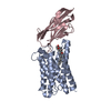 8apyC 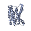 6zxrS S: Starting model for refinement C: citing same article ( |
|---|---|
| Similar structure data |
-Links
 PDBj
PDBj


- Assembly
| Deposited unit | 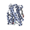
| ||||||||
|---|---|---|---|---|---|---|---|---|---|
| 1 |
| ||||||||
| Unit cell |
|
-Components
| #1: Protein | Mass: 24476.889 Da / Num. of mol.: 1 Source method: isolated from a genetically manipulated source Source: (gene. exp.)  | ||||||
|---|---|---|---|---|---|---|---|
| #2: Protein/peptide | Mass: 848.833 Da / Num. of mol.: 1 / Source method: obtained synthetically / Details: peptide / Source: (synth.) Homo sapiens (human) | ||||||
| #3: Chemical | ChemComp-OLC / (   Mass: 356.540 Da / Num. of mol.: 4 / Source method: obtained synthetically / Formula: C21H40O4 Mass: 356.540 Da / Num. of mol.: 4 / Source method: obtained synthetically / Formula: C21H40O4#4: Water | ChemComp-HOH / |  Mass: 18.015 Da / Num. of mol.: 51 / Source method: isolated from a natural source / Formula: H2O Mass: 18.015 Da / Num. of mol.: 51 / Source method: isolated from a natural source / Formula: H2OHas ligand of interest | N | Has protein modification | N | |
-Experimental details
-Experiment
| Experiment | Method: X-RAY DIFFRACTION / Number of used crystals: 1 |
|---|
- Sample preparation
| Crystal | Density Matthews: 2.19 Å3/Da / Density % sol: 43.88 % |
|---|---|
| Crystal grow | Temperature: 292 K / Method: lipidic cubic phase / pH: 6 / Details: 30% PEG 600, 0.1M MES pH 6.0, 0.1M Sodium Nitrate |
-Data collection
| Diffraction | Mean temperature: 100 K / Serial crystal experiment: N | ||||||||||||||||||||||||
|---|---|---|---|---|---|---|---|---|---|---|---|---|---|---|---|---|---|---|---|---|---|---|---|---|---|
| Diffraction source | Source: SYNCHROTRON / Site: Diamond / Beamline: I24 / Wavelength: 0.9999 Å | ||||||||||||||||||||||||
| Detector | Type: DECTRIS PILATUS3 6M / Detector: PIXEL / Date: Oct 6, 2020 | ||||||||||||||||||||||||
| Radiation | Protocol: SINGLE WAVELENGTH / Monochromatic (M) / Laue (L): M / Scattering type: x-ray | ||||||||||||||||||||||||
| Radiation wavelength | Wavelength: 0.9999 Å / Relative weight: 1 | ||||||||||||||||||||||||
| Reflection | Resolution: 2.283→62.28 Å / Num. obs: 10122 / % possible obs: 99.3 % / Redundancy: 3.1 % / Biso Wilson estimate: 37.5 Å2 / Rpim(I) all: 0.159 / Rrim(I) all: 0.289 / Net I/σ(I): 5.1 | ||||||||||||||||||||||||
| Reflection shell | Diffraction-ID: 1
|
- Processing
| Software |
| ||||||||||||||||||||||||||||||||||||||||||||||||||||||||||||||||||||||||||||||||||||||||||||||||||||||||||||
|---|---|---|---|---|---|---|---|---|---|---|---|---|---|---|---|---|---|---|---|---|---|---|---|---|---|---|---|---|---|---|---|---|---|---|---|---|---|---|---|---|---|---|---|---|---|---|---|---|---|---|---|---|---|---|---|---|---|---|---|---|---|---|---|---|---|---|---|---|---|---|---|---|---|---|---|---|---|---|---|---|---|---|---|---|---|---|---|---|---|---|---|---|---|---|---|---|---|---|---|---|---|---|---|---|---|---|---|---|---|
| Refinement | Method to determine structure: MOLECULAR REPLACEMENT Starting model: 6ZXR Resolution: 2.283→39.41 Å / Cor.coef. Fo:Fc: 0.908 / Cor.coef. Fo:Fc free: 0.859 / SU R Cruickshank DPI: 0.584 / Cross valid method: THROUGHOUT / σ(F): 0 / SU R Blow DPI: 0.488 / SU Rfree Blow DPI: 0.281 / SU Rfree Cruickshank DPI: 0.287 Details: HYDROGENS WERE FULLY REFINED WITH ZERO OCCUPANCY AT NUCLEAR POSITION. REFINEMENT NOTES. NUMBER OF REFINEMENT NOTES : 1 NOTE 1 : IDEAL-DIST CONTACT TERM CONTACT SETUP. ALL ATOMS HAVE CCP4 ATOM TYPE FROM LIBRARY
| ||||||||||||||||||||||||||||||||||||||||||||||||||||||||||||||||||||||||||||||||||||||||||||||||||||||||||||
| Displacement parameters | Biso max: 88.58 Å2 / Biso mean: 39.63 Å2 / Biso min: 14.66 Å2
| ||||||||||||||||||||||||||||||||||||||||||||||||||||||||||||||||||||||||||||||||||||||||||||||||||||||||||||
| Refine analyze | Luzzati coordinate error obs: 0.38 Å | ||||||||||||||||||||||||||||||||||||||||||||||||||||||||||||||||||||||||||||||||||||||||||||||||||||||||||||
| Refinement step | Cycle: final / Resolution: 2.283→39.41 Å
| ||||||||||||||||||||||||||||||||||||||||||||||||||||||||||||||||||||||||||||||||||||||||||||||||||||||||||||
| Refine LS restraints |
| ||||||||||||||||||||||||||||||||||||||||||||||||||||||||||||||||||||||||||||||||||||||||||||||||||||||||||||
| LS refinement shell | Resolution: 2.283→2.31 Å / Rfactor Rfree error: 0
|
