 Movie
Movie Controller


+ Open data

- Basic information
| Entry | Database: PDB / ID: 8aim | ||||||
|---|---|---|---|---|---|---|---|
| Title | Ugi-2 SAUNG complex | ||||||
 Components Components |
| ||||||
 Keywords Keywords | PROTEIN BINDING / Ugi / Ung / complex / inhibitor | ||||||
| Function / homology |  Function and homology information Function and homology informationbase-excision repair, AP site formation via deaminated base removal / uracil-DNA glycosylase / uracil DNA N-glycosylase activity / cytoplasm Similarity search - Function | ||||||
| Biological species |  Bacillus phage vB_BpuM-BpSp (virus) Bacillus phage vB_BpuM-BpSp (virus)  Staphylococcus aureus (bacteria) Staphylococcus aureus (bacteria) | ||||||
| Method |  X-RAY DIFFRACTION / SYNCHROTRON / MOLECULAR REPLACEMENT / Resolution: 2.6 Å X-RAY DIFFRACTION / SYNCHROTRON / MOLECULAR REPLACEMENT / Resolution: 2.6 Å | ||||||
 Authors Authors | Muselmani, W. / Savva, R. | ||||||
| Funding support |  United Kingdom, 1items United Kingdom, 1items
| ||||||
 Citation Citation | Journal: Viruses / Year: 2023 Title: A Multimodal Approach towards Genomic Identification of Protein Inhibitors of Uracil-DNA Glycosylase. Authors: Muselmani, W. / Kashif-Khan, N. / Bagneris, C. / Santangelo, R. / Williams, M.A. / Savva, R. | ||||||
| History |
|
- Structure visualization
| Structure viewer | Molecule: MolmilJmol/JSmol |
|---|
- Downloads & links
-Download
| PDBx/mmCIF format | 8aim.cif.gz | 243.7 KB | Display | PDBx/mmCIF format |
|---|---|---|---|---|
| PDB format | pdb8aim.ent.gz | 193.5 KB | Display | PDB format |
| PDBx/mmJSON format | 8aim.json.gz | Tree view | PDBx/mmJSON format | |
| Others |  Other downloads Other downloads |
-Validation report
| Arichive directory | https://data.pdbj.org/pub/pdb/validation_reports/ai/8aimftp://data.pdbj.org/pub/pdb/validation_reports/ai/8aim | HTTPS FTP |
|---|
-Related structure data
| Related structure data |  8ailC 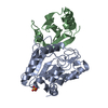 8ainC 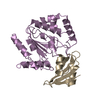 1udiS  3wdgS S: Starting model for refinement C: citing same article ( |
|---|---|
| Similar structure data |
-Links
 PDBj
PDBj- Assembly
| Deposited unit | 
| ||||||||||||||||||||||||||||||||||||||||||||||||||||||||||||||||||||||||||||
|---|---|---|---|---|---|---|---|---|---|---|---|---|---|---|---|---|---|---|---|---|---|---|---|---|---|---|---|---|---|---|---|---|---|---|---|---|---|---|---|---|---|---|---|---|---|---|---|---|---|---|---|---|---|---|---|---|---|---|---|---|---|---|---|---|---|---|---|---|---|---|---|---|---|---|---|---|---|
| 1 | 
| ||||||||||||||||||||||||||||||||||||||||||||||||||||||||||||||||||||||||||||
| 2 | 
| ||||||||||||||||||||||||||||||||||||||||||||||||||||||||||||||||||||||||||||
| Unit cell |
| ||||||||||||||||||||||||||||||||||||||||||||||||||||||||||||||||||||||||||||
| Noncrystallographic symmetry (NCS) | NCS domain:
NCS domain segments:
NCS ensembles :
|
-Components
| #1: Protein | Mass: 10427.682 Da / Num. of mol.: 2 Source method: isolated from a genetically manipulated source Source: (gene. exp.) Bacillus phage vB_BpuM-BpSp (virus) / Gene: Bp8pS_259 / Production host: #2: Protein | Mass: 26071.443 Da / Num. of mol.: 2 Source method: isolated from a genetically manipulated source Source: (gene. exp.) Staphylococcus aureus (bacteria)Gene: ung, A6762_02745, C7P97_05245, CSC87_03540, CV021_15850, E3A28_05745, E3K14_03025, GO782_00930, GO788_12235, NCTC6133_00684, NCTC7878_01215, NCTC7972_01353, QU38_09915, SAMEA70245418_01578 Production host: #3: Water | ChemComp-HOH / |  Mass: 18.015 Da / Num. of mol.: 49 / Source method: isolated from a natural source / Formula: H2O Mass: 18.015 Da / Num. of mol.: 49 / Source method: isolated from a natural source / Formula: H2OHas protein modification | Y | |
|---|
-Experimental details
-Experiment
| Experiment | Method: X-RAY DIFFRACTION / Number of used crystals: 1 |
|---|
- Sample preparation
| Crystal | Density Matthews: 2.79 Å3/Da / Density % sol: 55.85 % |
|---|---|
| Crystal grow | Temperature: 289.15 K / Method: vapor diffusion, sitting drop / Details: 0.1 M HEPES, pH 7.5, 70% v/v MPD |
-Data collection
| Diffraction | Mean temperature: 289.15 K / Serial crystal experiment: N |
|---|---|
| Diffraction source | Source: SYNCHROTRON / Site: Diamond / Beamline: I04 / Wavelength: 0.98 Å |
| Detector | Type: DECTRIS EIGER X 16M / Detector: PIXEL / Date: Oct 14, 2019 |
| Radiation | Protocol: SINGLE WAVELENGTH / Monochromatic (M) / Laue (L): M / Scattering type: x-ray |
| Radiation wavelength | Wavelength: 0.98 Å / Relative weight: 1 |
| Reflection | Resolution: 2.6→52.92 Å / Num. obs: 25462 / % possible obs: 100 % / Redundancy: 1.9 % / CC1/2: 0.98 / Net I/σ(I): 1.62 |
| Reflection shell | Resolution: 2.6→2.72 Å / Num. unique obs: 3052 / CC1/2: 0.528 |
- Processing
| Software |
| ||||||||||||||||||||||||||||||||||||||||||||||||||||||||||||||||||||||||||||||||||||||||||||||||||||||||||||||||||||||||||||||||||||||||||||||||||||||||||||||||
|---|---|---|---|---|---|---|---|---|---|---|---|---|---|---|---|---|---|---|---|---|---|---|---|---|---|---|---|---|---|---|---|---|---|---|---|---|---|---|---|---|---|---|---|---|---|---|---|---|---|---|---|---|---|---|---|---|---|---|---|---|---|---|---|---|---|---|---|---|---|---|---|---|---|---|---|---|---|---|---|---|---|---|---|---|---|---|---|---|---|---|---|---|---|---|---|---|---|---|---|---|---|---|---|---|---|---|---|---|---|---|---|---|---|---|---|---|---|---|---|---|---|---|---|---|---|---|---|---|---|---|---|---|---|---|---|---|---|---|---|---|---|---|---|---|---|---|---|---|---|---|---|---|---|---|---|---|---|---|---|---|---|
| Refinement | Method to determine structure: MOLECULAR REPLACEMENT Starting model: 3WDG, 1UDI Resolution: 2.6→52.92 Å / Cor.coef. Fo:Fc: 0.943 / Cor.coef. Fo:Fc free: 0.914 / SU B: 13.686 / SU ML: 0.268 / Cross valid method: FREE R-VALUE / ESU R: 0.564 / ESU R Free: 0.293 Details: Hydrogens have been added in their riding positions
| ||||||||||||||||||||||||||||||||||||||||||||||||||||||||||||||||||||||||||||||||||||||||||||||||||||||||||||||||||||||||||||||||||||||||||||||||||||||||||||||||
| Solvent computation | Ion probe radii: 0.8 Å / Shrinkage radii: 0.8 Å / VDW probe radii: 1.2 Å / Solvent model: MASK BULK SOLVENT | ||||||||||||||||||||||||||||||||||||||||||||||||||||||||||||||||||||||||||||||||||||||||||||||||||||||||||||||||||||||||||||||||||||||||||||||||||||||||||||||||
| Displacement parameters | Biso mean: 43.352 Å2
| ||||||||||||||||||||||||||||||||||||||||||||||||||||||||||||||||||||||||||||||||||||||||||||||||||||||||||||||||||||||||||||||||||||||||||||||||||||||||||||||||
| Refinement step | Cycle: LAST / Resolution: 2.6→52.92 Å
| ||||||||||||||||||||||||||||||||||||||||||||||||||||||||||||||||||||||||||||||||||||||||||||||||||||||||||||||||||||||||||||||||||||||||||||||||||||||||||||||||
| Refine LS restraints |
| ||||||||||||||||||||||||||||||||||||||||||||||||||||||||||||||||||||||||||||||||||||||||||||||||||||||||||||||||||||||||||||||||||||||||||||||||||||||||||||||||
| Refine LS restraints NCS |
| ||||||||||||||||||||||||||||||||||||||||||||||||||||||||||||||||||||||||||||||||||||||||||||||||||||||||||||||||||||||||||||||||||||||||||||||||||||||||||||||||
| LS refinement shell |
|
