 Movie
Movie Controller

[English] 日本語
 Yorodumi
- PDB-1udi: NUCLEOTIDE MIMICRY IN THE CRYSTAL STRUCTURE OF THE URACIL-DNA GLY... -
+ Open data

- Basic information
| Entry | Database: PDB / ID: 1udi | ||||||
|---|---|---|---|---|---|---|---|
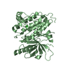
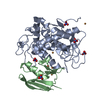
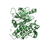
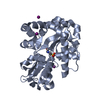
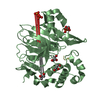
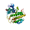
| Title | NUCLEOTIDE MIMICRY IN THE CRYSTAL STRUCTURE OF THE URACIL-DNA GLYCOSYLASE-URACIL GLYCOSYLASE INHIBITOR PROTEIN COMPLEX | ||||||
 Components Components |
| ||||||
 Keywords Keywords | HYDROLASE/INHIBITOR / HYDROLASE-INHIBITOR complex | ||||||
| Function / homology |  Function and homology information Function and homology informationbase-excision repair, AP site formation via deaminated base removal / uracil-DNA glycosylase / uracil DNA N-glycosylase activity / symbiont-mediated perturbation of host defense response / host cell nucleus Similarity search - Function | ||||||
| Biological species |  Herpes simplex virusBacillus phage PBS1 (virus) Herpes simplex virusBacillus phage PBS1 (virus) | ||||||
| Method |  X-RAY DIFFRACTION / SYNCHROTRON / Resolution: 2.7 Å X-RAY DIFFRACTION / SYNCHROTRON / Resolution: 2.7 Å | ||||||
 Authors Authors | Pearl, L.H. / Savva, R. | ||||||
 Citation Citation | Journal: Nat.Struct.Biol. / Year: 1995 Title: Nucleotide mimicry in the crystal structure of the uracil-DNA glycosylase-uracil glycosylase inhibitor protein complex. Authors: Savva, R. / Pearl, L.H. #1: Journal: Proteins / Year: 1995Title: Cloning and Expression of the Uracil-DNA Glycosylase Inhibitor from Bacteriophage Pbs1, and Crystallisation of a Uracil-DNA Glycosylase Uracil-DNA Glycosylase Inhibitor Complex Authors: Savva, R. / Pearl, L.H. #2: Journal: Nature / Year: 1995Title: The Structural Basis of Specific Base Excision Repair by Uracil-DNA Glycosylase Authors: Savva, R. / Mcauley-Hecht, K. / Brown, T. / Pearl, L.H. #3: Journal: J.Mol.Biol. / Year: 1993Title: Crystallization and Preliminary X-Ray Analysis of the Uracil-DNA Glycosylase DNA Repair Enzyme from Herpes Simplex Virus Type 1 Authors: Savva, R. / Pearl, L.H. | ||||||
| History |
|
- Structure visualization
| Structure viewer | Molecule: MolmilJmol/JSmol |
|---|
- Downloads & links
-Download
| PDBx/mmCIF format | 1udi.cif.gz | 72.8 KB | Display | PDBx/mmCIF format |
|---|---|---|---|---|
| PDB format | pdb1udi.ent.gz | 54.6 KB | Display | PDB format |
| PDBx/mmJSON format | 1udi.json.gz | Tree view | PDBx/mmJSON format | |
| Others |  Other downloads Other downloads |
-Validation report
| Arichive directory | https://data.pdbj.org/pub/pdb/validation_reports/ud/1udiftp://data.pdbj.org/pub/pdb/validation_reports/ud/1udi | HTTPS FTP |
|---|
-Related structure data
| Similar structure data |
|---|


-Links
 PDBj
PDBj- Assembly
| Deposited unit | 
| ||||||||
|---|---|---|---|---|---|---|---|---|---|
| 1 |
| ||||||||
| Unit cell |
| ||||||||
| Atom site foot note | 1: CIS PROLINE - PRO E 63 / 2: CIS PROLINE - PRO I 63 |
-Components
| #1: Protein | Mass: 27366.447 Da / Num. of mol.: 1 / Source method: isolated from a natural source / Source: (natural) Herpes simplex virus (type 1 / strain 17) / Genus: Simplexvirus / Species: Human herpesvirus 1 / Strain: 17 / References: UniProt: P10186, uridine nucleosidase |
|---|---|
| #2: Protein | Mass: 9351.478 Da / Num. of mol.: 1 / Source method: isolated from a natural source / Source: (natural) Bacillus phage PBS1 (virus) / References: UniProt: P14739 |
| #3: Water | ChemComp-HOH /  Mass: 18.015 Da / Num. of mol.: 47 / Source method: isolated from a natural source / Formula: H2O Mass: 18.015 Da / Num. of mol.: 47 / Source method: isolated from a natural source / Formula: H2O |
-Experimental details
-Experiment
| Experiment | Method: X-RAY DIFFRACTION / Number of used crystals: 1 |
|---|
- Sample preparation
| Crystal | Density Matthews: 3.34 Å3/Da / Density % sol: 63.19 % | |||||||||||||||||||||||||
|---|---|---|---|---|---|---|---|---|---|---|---|---|---|---|---|---|---|---|---|---|---|---|---|---|---|---|
| Crystal grow | *PLUS pH: 6.8 / Method: batch method | |||||||||||||||||||||||||
| Components of the solutions | *PLUS
|
-Data collection
| Diffraction source | Source: SYNCHROTRON / Site: SRS  / Beamline: PX7.2 / Wavelength: 1.448 / Beamline: PX7.2 / Wavelength: 1.448 |
|---|---|
| Detector | Type: MAR scanner 180 mm plate / Detector: IMAGE PLATE |
| Radiation | Monochromatic (M) / Laue (L): M / Scattering type: x-ray |
| Radiation wavelength | Wavelength: 1.448 Å / Relative weight: 1 |
| Reflection | Num. obs: 12683 / % possible obs: 92.7 % / Redundancy: 2.3 % / Rmerge(I) obs: 0.07 |
| Reflection | *PLUS Rmerge(I) obs: 0.07 |
| Reflection shell | *PLUS Highest resolution: 2.7 Å / Lowest resolution: 2.77 Å / % possible obs: 85 % |
- Processing
| Software |
| ||||||||||||||||||||||||||||||||||||||||||||||||||||||||||||
|---|---|---|---|---|---|---|---|---|---|---|---|---|---|---|---|---|---|---|---|---|---|---|---|---|---|---|---|---|---|---|---|---|---|---|---|---|---|---|---|---|---|---|---|---|---|---|---|---|---|---|---|---|---|---|---|---|---|---|---|---|---|
| Refinement | Resolution: 2.7→8 Å / σ(F): 0
| ||||||||||||||||||||||||||||||||||||||||||||||||||||||||||||
| Refinement step | Cycle: LAST / Resolution: 2.7→8 Å
| ||||||||||||||||||||||||||||||||||||||||||||||||||||||||||||
| Refine LS restraints |
| ||||||||||||||||||||||||||||||||||||||||||||||||||||||||||||
| Software | *PLUS Name: X-PLOR / Classification: refinement | ||||||||||||||||||||||||||||||||||||||||||||||||||||||||||||
| Refinement | *PLUS | ||||||||||||||||||||||||||||||||||||||||||||||||||||||||||||
| Solvent computation | *PLUS | ||||||||||||||||||||||||||||||||||||||||||||||||||||||||||||
| Displacement parameters | *PLUS | ||||||||||||||||||||||||||||||||||||||||||||||||||||||||||||
| Refine LS restraints | *PLUS Type: x_improper_angle_deg / Dev ideal: 1.468 |
