 Movie
Movie Controller

[English] 日本語
 Yorodumi
- PDB-8agk: Botulinum neurotoxin subtype A6 cell binding domain in complex wi... -
+ Open data

- Basic information
| Entry | Database: PDB / ID: 8agk | ||||||
|---|---|---|---|---|---|---|---|
| Title | Botulinum neurotoxin subtype A6 cell binding domain in complex with GD1a ganglioside | ||||||
 Components Components | Bont/A1 | ||||||
 Keywords Keywords | TOXIN / Botulism / ganglioside / botulinum neurotoxin / cell binding domain / complex / subtype | ||||||
| Function / homology |  Function and homology information Function and homology informationprotein transmembrane transporter activity / metalloendopeptidase activity / toxin activity / proteolysis / extracellular region / zinc ion binding Similarity search - Function | ||||||
| Biological species |  Clostridium botulinum str. Iwanei E (bacteria) Clostridium botulinum str. Iwanei E (bacteria) | ||||||
| Method |  X-RAY DIFFRACTION / SYNCHROTRON / MOLECULAR REPLACEMENT / Resolution: 1.5 Å X-RAY DIFFRACTION / SYNCHROTRON / MOLECULAR REPLACEMENT / Resolution: 1.5 Å | ||||||
 Authors Authors | Gregory, K.S. / Acharya, K.R. / Liu, S.M. / Newell, A.R. / Mojanaga, O.O. | ||||||
| Funding support | 1items
| ||||||
 Citation Citation | Journal: Int J Mol Sci / Year: 2022 Title: Crystal Structures of the Clostridium botulinum Neurotoxin A6 Cell Binding Domain Alone and in Complex with GD1a Reveal Significant Conformational Flexibility. Authors: Gregory, K.S. / Newell, A.R. / Mojanaga, O.O. / Liu, S.M. / Acharya, K.R. | ||||||
| History |
|
- Structure visualization
| Structure viewer | Molecule: MolmilJmol/JSmol |
|---|
- Downloads & links
-Download
| PDBx/mmCIF format | 8agk.cif.gz | 114.5 KB | Display | PDBx/mmCIF format |
|---|---|---|---|---|
| PDB format | pdb8agk.ent.gz | Display | PDB format | |
| PDBx/mmJSON format | 8agk.json.gz | Tree view | PDBx/mmJSON format | |
| Others |  Other downloads Other downloads |
-Validation report
| Arichive directory | https://data.pdbj.org/pub/pdb/validation_reports/ag/8agkftp://data.pdbj.org/pub/pdb/validation_reports/ag/8agk | HTTPS FTP |
|---|
-Related structure data
| Related structure data | 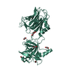 8alpC 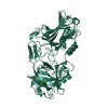 6twoS S: Starting model for refinement C: citing same article ( |
|---|---|
| Similar structure data |
-Links
 PDBj
PDBj- Assembly
| Deposited unit | 
| ||||||||
|---|---|---|---|---|---|---|---|---|---|
| 1 |
| ||||||||
| Unit cell |
|
-Components
| #1: Protein | Mass: 50681.645 Da / Num. of mol.: 1 Source method: isolated from a genetically manipulated source Source: (gene. exp.) Clostridium botulinum str. Iwanei E (bacteria)Production host: | ||||||||
|---|---|---|---|---|---|---|---|---|---|
| #2: Polysaccharide | N-acetyl-alpha-neuraminic acid-(2-3)-beta-D-galactopyranose-(1-3)-2-acetamido-2-deoxy-beta-D- ...N-acetyl-alpha-neuraminic acid-(2-3)-beta-D-galactopyranose-(1-3)-2-acetamido-2-deoxy-beta-D-galactopyranose-(1-4)-alpha-D-galactopyranose-(1-4)-alpha-D-glucopyranose Type: oligosaccharide / Mass: 998.885 Da / Num. of mol.: 1 Source method: isolated from a genetically manipulated source | ||||||||
| #3: Chemical | ChemComp-EDO /   Mass: 62.068 Da / Num. of mol.: 5 / Source method: obtained synthetically / Formula: C2H6O2 Mass: 62.068 Da / Num. of mol.: 5 / Source method: obtained synthetically / Formula: C2H6O2#4: Chemical |   Mass: 106.120 Da / Num. of mol.: 3 / Source method: obtained synthetically / Formula: C4H10O3 Mass: 106.120 Da / Num. of mol.: 3 / Source method: obtained synthetically / Formula: C4H10O3#5: Water | ChemComp-HOH / |  Mass: 18.015 Da / Num. of mol.: 197 / Source method: isolated from a natural source / Formula: H2O Mass: 18.015 Da / Num. of mol.: 197 / Source method: isolated from a natural source / Formula: H2OHas ligand of interest | Y | Has protein modification | Y | |
-Experimental details
-Experiment
| Experiment | Method: X-RAY DIFFRACTION / Number of used crystals: 1 |
|---|
- Sample preparation
| Crystal | Density Matthews: 2.15 Å3/Da / Density % sol: 42.74 % |
|---|---|
| Crystal grow | Temperature: 289.15 K / Method: vapor diffusion / Details: 0.2 M NaCl, 0.1 M HEPES pH 7.0, 20% w/v PEG 6000 |
-Data collection
| Diffraction | Mean temperature: 100 K / Serial crystal experiment: N |
|---|---|
| Diffraction source | Source: SYNCHROTRON / Site: Diamond  / Beamline: I04 / Wavelength: 0.9795 Å / Beamline: I04 / Wavelength: 0.9795 Å |
| Detector | Type: DECTRIS EIGER2 X 16M / Detector: PIXEL / Date: Oct 16, 2021 |
| Radiation | Protocol: SINGLE WAVELENGTH / Monochromatic (M) / Laue (L): M / Scattering type: x-ray |
| Radiation wavelength | Wavelength: 0.9795 Å / Relative weight: 1 |
| Reflection | Resolution: 1.5→57.65 Å / Num. obs: 67218 / % possible obs: 100 % / Redundancy: 13.6 % / CC1/2: 0.998 / Rpim(I) all: 0.05 / Net I/σ(I): 7.2 |
| Reflection shell | Resolution: 1.5→1.53 Å / Redundancy: 13.6 % / Mean I/σ(I) obs: 0.3 / Num. unique obs: 3357 / CC1/2: 0.991 / Rpim(I) all: 2.241 / % possible all: 100 |
- Processing
| Software |
| |||||||||||||||||||||||||||||||||||||||||||||||||||||||||||||||||||||||||||||||||||||||||||||||||||||||||||||||||||||||||||||||||||||||||||||||||||||||||||
|---|---|---|---|---|---|---|---|---|---|---|---|---|---|---|---|---|---|---|---|---|---|---|---|---|---|---|---|---|---|---|---|---|---|---|---|---|---|---|---|---|---|---|---|---|---|---|---|---|---|---|---|---|---|---|---|---|---|---|---|---|---|---|---|---|---|---|---|---|---|---|---|---|---|---|---|---|---|---|---|---|---|---|---|---|---|---|---|---|---|---|---|---|---|---|---|---|---|---|---|---|---|---|---|---|---|---|---|---|---|---|---|---|---|---|---|---|---|---|---|---|---|---|---|---|---|---|---|---|---|---|---|---|---|---|---|---|---|---|---|---|---|---|---|---|---|---|---|---|---|---|---|---|---|---|---|---|
| Refinement | Method to determine structure: MOLECULAR REPLACEMENT Starting model: 6TWO Resolution: 1.5→57.65 Å / Cor.coef. Fo:Fc: 0.967 / Cor.coef. Fo:Fc free: 0.957 / SU B: 3.207 / SU ML: 0.103 / Cross valid method: FREE R-VALUE / ESU R: 0.086 / ESU R Free: 0.087 Details: Hydrogens have been added in their riding positions
| |||||||||||||||||||||||||||||||||||||||||||||||||||||||||||||||||||||||||||||||||||||||||||||||||||||||||||||||||||||||||||||||||||||||||||||||||||||||||||
| Solvent computation | Ion probe radii: 0.8 Å / Shrinkage radii: 0.8 Å / VDW probe radii: 1.2 Å / Solvent model: MASK BULK SOLVENT | |||||||||||||||||||||||||||||||||||||||||||||||||||||||||||||||||||||||||||||||||||||||||||||||||||||||||||||||||||||||||||||||||||||||||||||||||||||||||||
| Displacement parameters | Biso mean: 27.134 Å2
| |||||||||||||||||||||||||||||||||||||||||||||||||||||||||||||||||||||||||||||||||||||||||||||||||||||||||||||||||||||||||||||||||||||||||||||||||||||||||||
| Refinement step | Cycle: LAST / Resolution: 1.5→57.65 Å
| |||||||||||||||||||||||||||||||||||||||||||||||||||||||||||||||||||||||||||||||||||||||||||||||||||||||||||||||||||||||||||||||||||||||||||||||||||||||||||
| Refine LS restraints |
| |||||||||||||||||||||||||||||||||||||||||||||||||||||||||||||||||||||||||||||||||||||||||||||||||||||||||||||||||||||||||||||||||||||||||||||||||||||||||||
| LS refinement shell |
|
