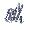
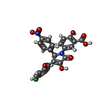
Entry Database : PDB / ID : 8a9gTitle Binary complex of 14-3-3 zeta Glucocorticoid Receptor (GR) pT524 peptide stabilised by (R)-para chloropyrrolidone1 14-3-3 protein zeta/delta Glucocorticoid receptor Keywords / / / / / / / / Function / homology Function Domain/homology Component
/ / / / / / / / / / / / / / / / / / / / / / / / / / / / / / / / / / / / / / / / / / / / / / / / / / / / / / / / / / / / / / / / / / / / / / / / / / / / / / / / / / / / / / / / / / / / / / / / / / / / / / / / / / / / / / / / / / / / / / / / / / / / / / / / / / / / / / / / / / / / / / / Biological species Homo sapiens (human)Method / / / Resolution : 1.961 Å Authors Munier, C.C. / Edman, K. / Perry, M.W.D. / Ottmann, C. Funding support European Union, 1items Organization Grant number Country European Commission 675179 European Union
Journal : J.Med.Chem. / Year : 2022Title : Designing Selective Drug-like Molecular Glues for the Glucocorticoid Receptor/14-3-3 Protein-Protein Interaction.Authors : Pallesen, J.S. / Munier, C.C. / Bosica, F. / Andrei, S.A. / Edman, K. / Gunnarsson, A. / La Sala, G. / Putra, O.D. / Srdanovic, S. / Wilson, A.J. / Wissler, L. / Ottmann, C. / Perry, M.W.D. / O'Mahony, G. History Deposition Jun 28, 2022 Deposition site / Processing site Revision 1.0 Dec 28, 2022 Provider / Type Revision 1.1 Jan 4, 2023 Group / Category / citation_authorItem _citation.journal_volume / _citation.page_first ... _citation.journal_volume / _citation.page_first / _citation.page_last / _citation_author.identifier_ORCID Revision 1.2 Jan 31, 2024 Group / Refinement descriptionCategory / chem_comp_bond / pdbx_initial_refinement_modelRevision 1.3 Nov 13, 2024 Group / Category / pdbx_modification_feature / Item
Show all Show less
 Movie
Movie Controller

 Yorodumi
 Open data
 Basic information
 Components
 Keywords
 Function and homology information
 Homo sapiens (human)
 X-RAY DIFFRACTION /
  Authors
 Citation
 Structure visualization
 Downloads & links
 Other downloads
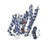

 PDBj
PDBj


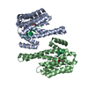
 Assembly
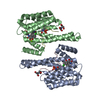


 Mass: 209.240 Da / Num. of mol.: 2 / Source method: obtained synthetically / Formula: C8H19NO5 / Comment: pH buffer*YM
 Mass: 494.838 Da / Num. of mol.: 2 / Source method: obtained synthetically / Formula: C24H15ClN2O8 / Feature type: SUBJECT OF INVESTIGATION
 Mass: 24.305 Da / Num. of mol.: 1 / Source method: obtained synthetically / Formula: Mg
 Sample preparation
 / Beamline: X06SA / Wavelength: 1.00003 Å
 Processing