 Movie
Movie Controller

[English] 日本語
 Yorodumi
- PDB-8a2q: Structure of the DNA-bound FANCD2-FANCI complex containing phosph... -
+ Open data

- Basic information
| Entry | Database: PDB / ID: 8a2q | ||||||||||||
|---|---|---|---|---|---|---|---|---|---|---|---|---|---|
| Title | Structure of the DNA-bound FANCD2-FANCI complex containing phosphomimetic FANCI | ||||||||||||
 Components Components |
| ||||||||||||
 Keywords Keywords | DNA BINDING PROTEIN / nucleic acid protein complex / DNA clamp / solenoid domains / Fanconi anemia / DNA repair / DNA damage / DNA inter strand crosslink / ubiquitination / ATR / pathway activation | ||||||||||||
| Function / homology |  Function and homology information Function and homology informationFanconi Anemia Pathway in DNA repair / Fanconi Anemia Pathway / Fanconi anaemia nuclear complex / homologous chromosome pairing at meiosis / double-strand break repair involved in meiotic recombination / mitotic intra-S DNA damage checkpoint signaling / interstrand cross-link repair / condensed chromosome / DNA polymerase binding / positive regulation of protein ubiquitination ...Fanconi Anemia Pathway in DNA repair / Fanconi Anemia Pathway / Fanconi anaemia nuclear complex / homologous chromosome pairing at meiosis / double-strand break repair involved in meiotic recombination / mitotic intra-S DNA damage checkpoint signaling / interstrand cross-link repair / condensed chromosome / DNA polymerase binding / positive regulation of protein ubiquitination / DNA repair / nucleoplasm / nucleus Similarity search - Function | ||||||||||||
| Biological species |  synthetic construct (others) | ||||||||||||
| Method | ELECTRON MICROSCOPY / single particle reconstruction / cryo EM / Resolution: 3.53 Å | ||||||||||||
 Authors Authors | Passmore, L.A. / Sijacki, T. / Alcon, P. | ||||||||||||
| Funding support |  United Kingdom, United Kingdom,  Germany, European Union, 3items Germany, European Union, 3items
| ||||||||||||
 Citation Citation | Journal: Nat Struct Mol Biol / Year: 2022 Title: The DNA-damage kinase ATR activates the FANCD2-FANCI clamp by priming it for ubiquitination. Authors: Tamara Sijacki / Pablo Alcón / Zhuo A Chen / Stephen H McLaughlin / Shabih Shakeel / Juri Rappsilber / Lori A Passmore /  Abstract: DNA interstrand cross-links are tumor-inducing lesions that block DNA replication and transcription. When cross-links are detected at stalled replication forks, ATR kinase phosphorylates FANCI, which ...DNA interstrand cross-links are tumor-inducing lesions that block DNA replication and transcription. When cross-links are detected at stalled replication forks, ATR kinase phosphorylates FANCI, which stimulates monoubiquitination of the FANCD2-FANCI clamp by the Fanconi anemia core complex. Monoubiquitinated FANCD2-FANCI is locked onto DNA and recruits nucleases that mediate DNA repair. However, it remains unclear how phosphorylation activates this pathway. Here, we report structures of FANCD2-FANCI complexes containing phosphomimetic FANCI. We observe that, unlike wild-type FANCD2-FANCI, the phosphomimetic complex closes around DNA, independent of the Fanconi anemia core complex. The phosphomimetic mutations do not substantially alter DNA binding but instead destabilize the open state of FANCD2-FANCI and alter its conformational dynamics. Overall, our results demonstrate that phosphorylation primes the FANCD2-FANCI clamp for ubiquitination, showing how multiple posttranslational modifications are coordinated to control DNA repair. | ||||||||||||
| History |
|
- Structure visualization
| Structure viewer | Molecule: MolmilJmol/JSmol |
|---|
- Downloads & links
-Download
| PDBx/mmCIF format | 8a2q.cif.gz | 635.2 KB | Display | PDBx/mmCIF format |
|---|---|---|---|---|
| PDB format | pdb8a2q.ent.gz | 492 KB | Display | PDB format |
| PDBx/mmJSON format | 8a2q.json.gz | Tree view | PDBx/mmJSON format | |
| Others |  Other downloads Other downloads |
-Validation report
| Arichive directory | https://data.pdbj.org/pub/pdb/validation_reports/a2/8a2qftp://data.pdbj.org/pub/pdb/validation_reports/a2/8a2q | HTTPS FTP |
|---|
-Related structure data
| Related structure data |  15101MC C: citing same article ( M: map data used to model this data |
|---|---|
| Similar structure data |


-Links
 PDBj
PDBj


- Assembly
| Deposited unit | 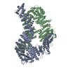
|
|---|---|
| 1 |
|
-Components
| #1: Protein | Mass: 164731.344 Da / Num. of mol.: 1 Source method: isolated from a genetically manipulated source Source: (gene. exp.)   Spodoptera frugiperda (fall armyworm) / References: UniProt: F1NP22 Spodoptera frugiperda (fall armyworm) / References: UniProt: F1NP22 |
|---|---|
| #2: Protein | Mass: 150357.156 Da / Num. of mol.: 1 Source method: isolated from a genetically manipulated source Source: (gene. exp.) Spodoptera frugiperda (fall armyworm) / References: UniProt: B0I564 |
| #3: DNA chain | Mass: 13554.697 Da / Num. of mol.: 1 / Source method: obtained synthetically / Source: (synth.) synthetic construct (others) |
| #4: DNA chain | Mass: 13541.715 Da / Num. of mol.: 1 / Source method: obtained synthetically / Source: (synth.) synthetic construct (others) |
-Experimental details
-Experiment
| Experiment | Method: ELECTRON MICROSCOPY |
|---|---|
| EM experiment | Aggregation state: PARTICLE / 3D reconstruction method: single particle reconstruction |
- Sample preparation
| Component |
| ||||||||||||||||||||||||||||
|---|---|---|---|---|---|---|---|---|---|---|---|---|---|---|---|---|---|---|---|---|---|---|---|---|---|---|---|---|---|
| Molecular weight |
| ||||||||||||||||||||||||||||
| Source (natural) |
| ||||||||||||||||||||||||||||
| Source (recombinant) |
| ||||||||||||||||||||||||||||
| Buffer solution | pH: 8 | ||||||||||||||||||||||||||||
| Buffer component |
| ||||||||||||||||||||||||||||
| Specimen | Conc.: 0.5 mg/ml / Embedding applied: NO / Shadowing applied: NO / Staining applied: NO / Vitrification applied: YES | ||||||||||||||||||||||||||||
| Specimen support | Grid material: GOLD / Grid type: UltrAuFoil R0.6/1 | ||||||||||||||||||||||||||||
| Vitrification | Instrument: FEI VITROBOT MARK IV / Cryogen name: ETHANE / Humidity: 100 % / Chamber temperature: 277.15 K |
- Electron microscopy imaging
| Experimental equipment |  Model: Titan Krios / Image courtesy: FEI Company |
|---|---|
| Microscopy | Model: FEI TITAN KRIOS |
| Electron gun | Electron source:  FIELD EMISSION GUN / Accelerating voltage: 300 kV / Illumination mode: FLOOD BEAM FIELD EMISSION GUN / Accelerating voltage: 300 kV / Illumination mode: FLOOD BEAM |
| Electron lens | Mode: BRIGHT FIELD / Nominal magnification: 105000 X / Nominal defocus max: 3000 nm / Nominal defocus min: 1500 nm / Cs: 2.7 mm / C2 aperture diameter: 50 µm / Alignment procedure: COMA FREE |
| Specimen holder | Cryogen: NITROGEN / Specimen holder model: FEI TITAN KRIOS AUTOGRID HOLDER |
| Image recording | Average exposure time: 1.5 sec. / Electron dose: 40 e/Å2 / Film or detector model: GATAN K3 (6k x 4k) / Num. of grids imaged: 1 / Num. of real images: 14842 |
| EM imaging optics | Energyfilter name: GIF Quantum LS / Energyfilter slit width: 20 eV |
- Processing
| EM software |
| ||||||||||||||||||||||||||||||||||||||||||||||||||||
|---|---|---|---|---|---|---|---|---|---|---|---|---|---|---|---|---|---|---|---|---|---|---|---|---|---|---|---|---|---|---|---|---|---|---|---|---|---|---|---|---|---|---|---|---|---|---|---|---|---|---|---|---|---|
| CTF correction | Type: PHASE FLIPPING AND AMPLITUDE CORRECTION | ||||||||||||||||||||||||||||||||||||||||||||||||||||
| Particle selection | Num. of particles selected: 1356378 | ||||||||||||||||||||||||||||||||||||||||||||||||||||
| Symmetry | Point symmetry: C1 (asymmetric) | ||||||||||||||||||||||||||||||||||||||||||||||||||||
| 3D reconstruction | Resolution: 3.53 Å / Resolution method: FSC 0.143 CUT-OFF / Num. of particles: 197436 / Num. of class averages: 1 / Symmetry type: POINT | ||||||||||||||||||||||||||||||||||||||||||||||||||||
| Atomic model building | Protocol: OTHER / Space: REAL Details: For dsDNA (chain S and T)an ideal B form DNA was generated and fitted into the density using the same softwares used to fit FANCD2 and FANCI chains (chain A and B) into the corresponding density. | ||||||||||||||||||||||||||||||||||||||||||||||||||||
| Atomic model building | 3D fitting-ID: 1 / Accession code: 6TNF / Initial refinement model-ID: 1 / PDB-ID: 6TNF / Source name: PDB / Type: experimental model
|

