 Movie
Movie Controller

[English] 日本語
 Yorodumi
- PDB-8a21: Crystal structure of Phosphoserine phosphatase SerB from Mycobact... -
+ Open data

- Basic information
| Entry | Database: PDB / ID: 8a21 | ||||||
|---|---|---|---|---|---|---|---|
| Title | Crystal structure of Phosphoserine phosphatase SerB from Mycobacterium avium in complex with phenylimidazole | ||||||
 Components Components | Phosphoserine phosphatase | ||||||
 Keywords Keywords | HYDROLASE / phosphoserine phosphatase / avium / PIM / phenylimidazole | ||||||
| Function / homology |  Function and homology information Function and homology informationphosphoserine phosphatase / L-phosphoserine phosphatase activity / L-serine biosynthetic process / metal ion binding Similarity search - Function | ||||||
| Biological species |  Mycobacterium avium 104 (bacteria) Mycobacterium avium 104 (bacteria) | ||||||
| Method |  X-RAY DIFFRACTION / SYNCHROTRON / MOLECULAR REPLACEMENT / molecular replacement / Resolution: 2.18 Å X-RAY DIFFRACTION / SYNCHROTRON / MOLECULAR REPLACEMENT / molecular replacement / Resolution: 2.18 Å | ||||||
 Authors Authors | Haufroid, M. / Wouters, J. | ||||||
| Funding support | 1items
| ||||||
 Citation Citation | Journal: Eur.J.Med.Chem. / Year: 2022 Title: Targeting the phosphoserine phosphatase MtSerB2 for tuberculosis drug discovery, an hybrid knowledge based /fragment based approach. Authors: Haufroid, M. / Volkov, A.N. / Wouters, J. | ||||||
| History |
|
- Structure visualization
| Structure viewer | Molecule: MolmilJmol/JSmol |
|---|
- Downloads & links
-Download
| PDBx/mmCIF format | 8a21.cif.gz | 93.3 KB | Display | PDBx/mmCIF format |
|---|---|---|---|---|
| PDB format | pdb8a21.ent.gz | 67.7 KB | Display | PDB format |
| PDBx/mmJSON format | 8a21.json.gz | Tree view | PDBx/mmJSON format | |
| Others |  Other downloads Other downloads |
-Validation report
| Arichive directory | https://data.pdbj.org/pub/pdb/validation_reports/a2/8a21ftp://data.pdbj.org/pub/pdb/validation_reports/a2/8a21 | HTTPS FTP |
|---|
-Related structure data
| Related structure data |  8a1zC  3p96S S: Starting model for refinement C: citing same article ( |
|---|---|
| Similar structure data |
-Links
 PDBj
PDBj

- Assembly
| Deposited unit | 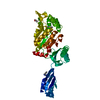
| ||||||||
|---|---|---|---|---|---|---|---|---|---|
| 1 | 
| ||||||||
| Unit cell |
|
-Components
-Protein , 1 types, 1 molecules A
| #1: Protein | Mass: 43852.094 Da / Num. of mol.: 1 Source method: isolated from a genetically manipulated source Source: (gene. exp.) Mycobacterium avium 104 (bacteria) / Gene: O972_03940 / Production host: |
|---|
-Non-polymers , 5 types, 216 molecules 


| #2: Chemical | ChemComp-MG /  Mass: 24.305 Da / Num. of mol.: 1 / Source method: obtained synthetically / Formula: Mg Mass: 24.305 Da / Num. of mol.: 1 / Source method: obtained synthetically / Formula: Mg | ||||||
|---|---|---|---|---|---|---|---|
| #3: Chemical |  Mass: 144.173 Da / Num. of mol.: 2 / Source method: obtained synthetically / Formula: C9H8N2 Mass: 144.173 Da / Num. of mol.: 2 / Source method: obtained synthetically / Formula: C9H8N2#4: Chemical | ChemComp-CL / |  Mass: 35.453 Da / Num. of mol.: 1 / Source method: obtained synthetically / Formula: Cl Mass: 35.453 Da / Num. of mol.: 1 / Source method: obtained synthetically / Formula: Cl#5: Chemical | ChemComp-GOL / |  Mass: 92.094 Da / Num. of mol.: 1 / Source method: obtained synthetically / Formula: C3H8O3 / Feature type: SUBJECT OF INVESTIGATION Mass: 92.094 Da / Num. of mol.: 1 / Source method: obtained synthetically / Formula: C3H8O3 / Feature type: SUBJECT OF INVESTIGATION#6: Water | ChemComp-HOH / | Mass: 18.015 Da / Num. of mol.: 211 / Source method: isolated from a natural source / Formula: H2O |
-Details
| Has ligand of interest | Y |
|---|
-Experimental details
-Experiment
| Experiment | Method: X-RAY DIFFRACTION / Number of used crystals: 1 |
|---|
- Sample preparation
| Crystal | Density Matthews: 2.57 Å3/Da / Density % sol: 52.2 % |
|---|---|
| Crystal grow | Temperature: 290 K / Method: vapor diffusion, hanging drop / pH: 6.1 / Details: MgCl2 0,5M; 20%PEG6000 MES 0.1M pH6.1 |
-Data collection
| Diffraction | Mean temperature: 100 K / Serial crystal experiment: N |
|---|---|
| Diffraction source | Source: SYNCHROTRON / Site: SOLEIL  / Beamline: PROXIMA 1 / Wavelength: 0.9786 Å / Beamline: PROXIMA 1 / Wavelength: 0.9786 Å |
| Detector | Type: DECTRIS EIGER X 16M / Detector: PIXEL / Date: Jun 1, 2018 |
| Radiation | Protocol: SINGLE WAVELENGTH / Monochromatic (M) / Laue (L): M / Scattering type: x-ray |
| Radiation wavelength | Wavelength: 0.9786 Å / Relative weight: 1 |
| Reflection | Resolution: 2.18→43.09 Å / Num. obs: 25224 / % possible obs: 98.76 % / Redundancy: 13.3 % / Biso Wilson estimate: 35.66 Å2 / CC1/2: 0.999 / CC star: 1 / Rmerge(I) obs: 0.1115 / Rpim(I) all: 0.0319 / Rrim(I) all: 0.1161 / Net I/σ(I): 16.22 |
| Reflection shell | Resolution: 2.18→2.26 Å / Redundancy: 11.6 % / Rmerge(I) obs: 1.531 / Mean I/σ(I) obs: 1.51 / Num. unique obs: 2238 / CC1/2: 0.777 / CC star: 0.935 / Rpim(I) all: 0.4595 / % possible all: 87.94 |
-Phasing
| Phasing | Method: molecular replacement |
|---|
- Processing
| Software |
| |||||||||||||||||||||||||||||||||||||||||||||||||||||||||||||||||||||||||||||||||||||||||||||||||||||||||
|---|---|---|---|---|---|---|---|---|---|---|---|---|---|---|---|---|---|---|---|---|---|---|---|---|---|---|---|---|---|---|---|---|---|---|---|---|---|---|---|---|---|---|---|---|---|---|---|---|---|---|---|---|---|---|---|---|---|---|---|---|---|---|---|---|---|---|---|---|---|---|---|---|---|---|---|---|---|---|---|---|---|---|---|---|---|---|---|---|---|---|---|---|---|---|---|---|---|---|---|---|---|---|---|---|---|---|
| Refinement | Method to determine structure: MOLECULAR REPLACEMENT Starting model: 3p96 Resolution: 2.18→43.09 Å / SU ML: 0.37 / Cross valid method: THROUGHOUT / σ(F): 1.34 / Phase error: 29.59 / Stereochemistry target values: ML
| |||||||||||||||||||||||||||||||||||||||||||||||||||||||||||||||||||||||||||||||||||||||||||||||||||||||||
| Solvent computation | Shrinkage radii: 0.9 Å / VDW probe radii: 1.11 Å / Solvent model: FLAT BULK SOLVENT MODEL | |||||||||||||||||||||||||||||||||||||||||||||||||||||||||||||||||||||||||||||||||||||||||||||||||||||||||
| Displacement parameters | Biso max: 69.82 Å2 / Biso mean: 37.0195 Å2 / Biso min: 16.97 Å2 | |||||||||||||||||||||||||||||||||||||||||||||||||||||||||||||||||||||||||||||||||||||||||||||||||||||||||
| Refinement step | Cycle: final / Resolution: 2.18→43.09 Å
| |||||||||||||||||||||||||||||||||||||||||||||||||||||||||||||||||||||||||||||||||||||||||||||||||||||||||
| LS refinement shell | Refine-ID: X-RAY DIFFRACTION / Rfactor Rfree error: 0 / Total num. of bins used: 14
|
