 Movie
Movie Controller


+ Open data

- Basic information
| Entry | Database: PDB / ID: 7ztk | ||||||
|---|---|---|---|---|---|---|---|
| Title | NME1 in complex with CoA | ||||||
 Components Components | Nucleoside diphosphate kinase A | ||||||
 Keywords Keywords | TRANSFERASE / NME1 / complex / nucleoside diphosphate kinase 1 / coenzyme-A | ||||||
| Function / homology |  Function and homology information Function and homology informationintermediate filament binding / negative regulation of myeloid leukocyte differentiation / Azathioprine ADME / Ribavirin ADME / farnesyl-diphosphate kinase / farnesyl diphosphate kinase activity / succinyl-CoA binding / ITP biosynthetic process / Interconversion of nucleotide di- and triphosphates / isoprenoid metabolic process ...intermediate filament binding / negative regulation of myeloid leukocyte differentiation / Azathioprine ADME / Ribavirin ADME / farnesyl-diphosphate kinase / farnesyl diphosphate kinase activity / succinyl-CoA binding / ITP biosynthetic process / Interconversion of nucleotide di- and triphosphates / isoprenoid metabolic process / Hydrolases; Acting on ester bonds; Site specific endodeoxyribonucleases: cleavage is not sequence specific (deleted sub-subclass) / phosphotransferase activity, phosphate group as acceptor / DNA nuclease activity / acetyl-CoA catabolic process / nucleoside triphosphate biosynthetic process / acetyl-CoA binding / nucleoside diphosphate metabolic process / coenzyme A binding / protein histidine kinase activity / regulation of fatty acid biosynthetic process / apoptotic DNA fragmentation / nucleoside-diphosphate kinase / 3'-5'-DNA exonuclease activity / mammary gland development / UTP biosynthetic process / CTP biosynthetic process / gamma-tubulin binding / protein hexamerization / DNA catabolic process / nucleoside diphosphate kinase activity / dTMP biosynthetic process / GTP biosynthetic process / intermediate filament / Hydrolases; Acting on ester bonds; Exodeoxyribonucleases producing 5'-phosphomonoesters / histidine kinase / ribosomal small subunit binding / lactation / positive regulation of epithelial cell proliferation / RNA polymerase II transcription regulatory region sequence-specific DNA binding / DNA endonuclease activity / positive regulation of neuron projection development / ADP binding / ruffle membrane / endocytosis / kinase activity / GDP binding / myelin sheath / nervous system development / single-stranded DNA binding / early endosome / cell differentiation / mitochondrial outer membrane / non-specific serine/threonine protein kinase / negative regulation of gene expression / centrosome / protein kinase binding / perinuclear region of cytoplasm / magnesium ion binding / enzyme binding / mitochondrion / DNA binding / ATP binding / identical protein binding / nucleus / cytoplasm / cytosol Similarity search - Function | ||||||
| Biological species |  | ||||||
| Method |  X-RAY DIFFRACTION / SYNCHROTRON / MOLECULAR REPLACEMENT / Resolution: 2.6 Å X-RAY DIFFRACTION / SYNCHROTRON / MOLECULAR REPLACEMENT / Resolution: 2.6 Å | ||||||
 Authors Authors | Garcia-Saez, I. / Iuso, D. / Khochbin, S. / Petosa, C. | ||||||
| Funding support |  France, 1items France, 1items
| ||||||
 Citation Citation | Journal: Sci Adv / Year: 2023 Title: Nucleoside diphosphate kinases 1 and 2 regulate a protective liver response to a high-fat diet. Authors: Iuso, D. / Garcia-Saez, I. / Coute, Y. / Yamaryo-Botte, Y. / Boeri Erba, E. / Adrait, A. / Zeaiter, N. / Tokarska-Schlattner, M. / Jilkova, Z.M. / Boussouar, F. / Barral, S. / Signor, L. / ...Authors: Iuso, D. / Garcia-Saez, I. / Coute, Y. / Yamaryo-Botte, Y. / Boeri Erba, E. / Adrait, A. / Zeaiter, N. / Tokarska-Schlattner, M. / Jilkova, Z.M. / Boussouar, F. / Barral, S. / Signor, L. / Couturier, K. / Hajmirza, A. / Chuffart, F. / Bourova-Flin, E. / Vitte, A.L. / Bargier, L. / Puthier, D. / Decaens, T. / Rousseaux, S. / Botte, C. / Schlattner, U. / Petosa, C. / Khochbin, S. | ||||||
| History |
|
- Structure visualization
| Structure viewer | Molecule: MolmilJmol/JSmol |
|---|
- Downloads & links
-Download
| PDBx/mmCIF format | 7ztk.cif.gz | 374.6 KB | Display | PDBx/mmCIF format |
|---|---|---|---|---|
| PDB format | pdb7ztk.ent.gz | 307.9 KB | Display | PDB format |
| PDBx/mmJSON format | 7ztk.json.gz | Tree view | PDBx/mmJSON format | |
| Others |  Other downloads Other downloads |
-Validation report
| Arichive directory | https://data.pdbj.org/pub/pdb/validation_reports/zt/7ztkftp://data.pdbj.org/pub/pdb/validation_reports/zt/7ztk | HTTPS FTP |
|---|
-Related structure data
| Related structure data | 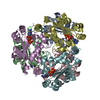 7zl8C  7zlwC 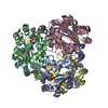 5ui4S C: citing same article ( S: Starting model for refinement |
|---|---|
| Similar structure data |
-Links
 PDBj
PDBj


- Assembly
| Deposited unit | 
| ||||||||
|---|---|---|---|---|---|---|---|---|---|
| 1 |
| ||||||||
| Unit cell |
|
-Components
| #1: Protein | Mass: 17561.199 Da / Num. of mol.: 6 Source method: isolated from a genetically manipulated source Details: NME1 hexamer in complex with CoA / Source: (gene. exp.) Production host:  References: UniProt: P15532, nucleoside-diphosphate kinase #2: Chemical | ChemComp-COA /   Mass: 767.534 Da / Num. of mol.: 6 / Source method: obtained synthetically / Formula: C21H36N7O16P3S / Feature type: SUBJECT OF INVESTIGATION Mass: 767.534 Da / Num. of mol.: 6 / Source method: obtained synthetically / Formula: C21H36N7O16P3S / Feature type: SUBJECT OF INVESTIGATION#3: Water | ChemComp-HOH / |  Mass: 18.015 Da / Num. of mol.: 77 / Source method: isolated from a natural source / Formula: H2O Mass: 18.015 Da / Num. of mol.: 77 / Source method: isolated from a natural source / Formula: H2OHas ligand of interest | Y | Has protein modification | N | |
|---|
-Experimental details
-Experiment
| Experiment | Method: X-RAY DIFFRACTION / Number of used crystals: 1 |
|---|
- Sample preparation
| Crystal | Density Matthews: 2.08 Å3/Da / Density % sol: 41 % |
|---|---|
| Crystal grow | Temperature: 293.15 K / Method: vapor diffusion, hanging drop / Details: 36% PEG 3350, 0.2M Mg(NO3) |
-Data collection
| Diffraction | Mean temperature: 100 K / Serial crystal experiment: N |
|---|---|
| Diffraction source | Source: SYNCHROTRON / Site: ESRF / Beamline: MASSIF-1 / Wavelength: 0.966 Å |
| Detector | Type: DECTRIS PILATUS3 2M / Detector: PIXEL / Date: Nov 17, 2018 |
| Radiation | Protocol: SINGLE WAVELENGTH / Monochromatic (M) / Laue (L): M / Scattering type: x-ray |
| Radiation wavelength | Wavelength: 0.966 Å / Relative weight: 1 |
| Reflection | Resolution: 2.6→49.13 Å / Num. obs: 26764 / % possible obs: 99.23 % / Redundancy: 3.2 % / Biso Wilson estimate: 40.72 Å2 / CC1/2: 0.945 / CC star: 0.986 / Rmerge(I) obs: 0.254 / Rpim(I) all: 0.17 / Rrim(I) all: 0.307 / Net I/σ(I): 3.37 |
| Reflection shell | Resolution: 2.6→2.693 Å / Redundancy: 3.3 % / Rmerge(I) obs: 1.046 / Mean I/σ(I) obs: 0.84 / Num. unique obs: 2649 / CC1/2: 0.542 / CC star: 0.839 / Rpim(I) all: 0.684 / Rrim(I) all: 1.253 / % possible all: 98.53 |
- Processing
| Software |
| ||||||||||||||||||||||||||||||||||||||||||||||||||||||||||||||||||||||
|---|---|---|---|---|---|---|---|---|---|---|---|---|---|---|---|---|---|---|---|---|---|---|---|---|---|---|---|---|---|---|---|---|---|---|---|---|---|---|---|---|---|---|---|---|---|---|---|---|---|---|---|---|---|---|---|---|---|---|---|---|---|---|---|---|---|---|---|---|---|---|---|
| Refinement | Method to determine structure: MOLECULAR REPLACEMENT Starting model: 5ui4 Resolution: 2.6→49.13 Å / SU ML: 0.4 / Cross valid method: THROUGHOUT / σ(F): 1.34 / Phase error: 31.35 / Stereochemistry target values: ML Details: TLS was used in the first runs of refinement (1 group/chain), but not in the last runs of refinement.
| ||||||||||||||||||||||||||||||||||||||||||||||||||||||||||||||||||||||
| Solvent computation | Shrinkage radii: 0.9 Å / VDW probe radii: 1.11 Å / Solvent model: FLAT BULK SOLVENT MODEL | ||||||||||||||||||||||||||||||||||||||||||||||||||||||||||||||||||||||
| Displacement parameters | Biso max: 131.33 Å2 / Biso mean: 47.6645 Å2 / Biso min: 18.25 Å2 | ||||||||||||||||||||||||||||||||||||||||||||||||||||||||||||||||||||||
| Refinement step | Cycle: final / Resolution: 2.6→49.13 Å
| ||||||||||||||||||||||||||||||||||||||||||||||||||||||||||||||||||||||
| LS refinement shell | Refine-ID: X-RAY DIFFRACTION / Rfactor Rfree error: 0 / Total num. of bins used: 9
|
