 Movie
Movie Controller


+ Open data

- Basic information
| Entry | Database: PDB / ID: 7zsr | ||||||
|---|---|---|---|---|---|---|---|
| Title | purine nucleoside phosphorylase in complex with JS-379 | ||||||
 Components Components | Purine nucleoside phosphorylase | ||||||
 Keywords Keywords | TRANSFERASE / PNP-inhibitor complex | ||||||
| Function / homology |  Function and homology information Function and homology informationnucleoside metabolic process / purine-nucleoside phosphorylase / purine-nucleoside phosphorylase activity / cytoplasm Similarity search - Function | ||||||
| Biological species |   Mycobacterium tuberculosis (bacteria) Mycobacterium tuberculosis (bacteria) | ||||||
| Method |  X-RAY DIFFRACTION / SYNCHROTRON / MOLECULAR REPLACEMENT / Resolution: 1.97 Å X-RAY DIFFRACTION / SYNCHROTRON / MOLECULAR REPLACEMENT / Resolution: 1.97 Å | ||||||
 Authors Authors | Djukic, S. / Pachl, P. / Rezacova, P. | ||||||
| Funding support | European Union, 1items
| ||||||
 Citation Citation | Journal: J.Med.Chem. / Year: 2023 Title: Design, Synthesis, Biological Evaluation, and Crystallographic Study of Novel Purine Nucleoside Phosphorylase Inhibitors. Authors: Skacel, J. / Djukic, S. / Baszczynski, O. / Kalcic, F. / Bilek, T. / Chalupsky, K. / Kozak, J. / Dvorakova, A. / Tloust'ova, E. / Kral'ova, Z. / Smidkova, M. / Voldrich, J. / Rumlova, M. / ...Authors: Skacel, J. / Djukic, S. / Baszczynski, O. / Kalcic, F. / Bilek, T. / Chalupsky, K. / Kozak, J. / Dvorakova, A. / Tloust'ova, E. / Kral'ova, Z. / Smidkova, M. / Voldrich, J. / Rumlova, M. / Pachl, P. / Brynda, J. / Vuckova, T. / Fabry, M. / Snasel, J. / Pichova, I. / Rezacova, P. / Mertlikova-Kaiserova, H. / Janeba, Z. | ||||||
| History |
|
- Structure visualization
| Structure viewer | Molecule: MolmilJmol/JSmol |
|---|
- Downloads & links
-Download
| PDBx/mmCIF format | 7zsr.cif.gz | 158.2 KB | Display | PDBx/mmCIF format |
|---|---|---|---|---|
| PDB format | pdb7zsr.ent.gz | 124.4 KB | Display | PDB format |
| PDBx/mmJSON format | 7zsr.json.gz | Tree view | PDBx/mmJSON format | |
| Others |  Other downloads Other downloads |
-Validation report
| Arichive directory | https://data.pdbj.org/pub/pdb/validation_reports/zs/7zsrftp://data.pdbj.org/pub/pdb/validation_reports/zs/7zsr | HTTPS FTP |
|---|
-Related structure data
| Related structure data |  7zslC  7zsmC  7zsnC 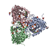 7zsoC 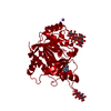 7zspC  7zsqC  8c25C 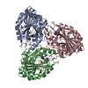 1g2oS S: Starting model for refinement C: citing same article ( |
|---|---|
| Similar structure data |
-Links
 PDBj
PDBj- Assembly
| Deposited unit | 
| ||||||||
|---|---|---|---|---|---|---|---|---|---|
| 1 |
| ||||||||
| Unit cell |
|
-Components
| #1: Protein | Mass: 27567.391 Da / Num. of mol.: 3 Source method: isolated from a genetically manipulated source Source: (gene. exp.) Mycobacterium tuberculosis (bacteria)Gene: deoD, E5M05_03615, E5M23_14660, E5M52_18960, E5M78_19105, ERS013440_01955, ERS027646_00621, ERS027659_03654, SAMEA2683035_02840 Production host: References: UniProt: A0A045IAS2, purine-nucleoside phosphorylase #2: Chemical |   Mass: 428.198 Da / Num. of mol.: 3 / Source method: obtained synthetically / Formula: C14H11BrN3O4PS / Feature type: SUBJECT OF INVESTIGATION Mass: 428.198 Da / Num. of mol.: 3 / Source method: obtained synthetically / Formula: C14H11BrN3O4PS / Feature type: SUBJECT OF INVESTIGATION#3: Chemical |   Mass: 59.044 Da / Num. of mol.: 3 / Source method: obtained synthetically / Formula: C2H3O2 Mass: 59.044 Da / Num. of mol.: 3 / Source method: obtained synthetically / Formula: C2H3O2#4: Water | ChemComp-HOH / |  Mass: 18.015 Da / Num. of mol.: 253 / Source method: isolated from a natural source / Formula: H2O Mass: 18.015 Da / Num. of mol.: 253 / Source method: isolated from a natural source / Formula: H2OHas ligand of interest | Y | |
|---|
-Experimental details
-Experiment
| Experiment | Method: X-RAY DIFFRACTION / Number of used crystals: 1 |
|---|
- Sample preparation
| Crystal | Density Matthews: 2.42 Å3/Da / Density % sol: 49.11 % |
|---|---|
| Crystal grow | Temperature: 291.15 K / Method: vapor diffusion, hanging drop Details: 0.1 M Tris; 25 mM Magnesium chloride; 25% w/v PEG4000; pH 7.5 |
-Data collection
| Diffraction | Mean temperature: 100 K / Serial crystal experiment: N |
|---|---|
| Diffraction source | Source: SYNCHROTRON / Site: BESSY  / Beamline: 14.2 / Wavelength: 0.918 Å / Beamline: 14.2 / Wavelength: 0.918 Å |
| Detector | Type: DECTRIS PILATUS 6M / Detector: PIXEL / Date: Nov 14, 2019 |
| Radiation | Protocol: SINGLE WAVELENGTH / Monochromatic (M) / Laue (L): M / Scattering type: x-ray |
| Radiation wavelength | Wavelength: 0.918 Å / Relative weight: 1 |
| Reflection | Resolution: 1.9→45.92 Å / Num. obs: 58542 / % possible obs: 81.5 % / Redundancy: 3.8 % / CC1/2: 0.996 / Net I/σ(I): 7.15 |
| Reflection shell | Resolution: 1.9→1.95 Å / Num. unique obs: 3729 / CC1/2: 0.44 |
- Processing
| Software |
| ||||||||||||||||||||||||||||||||||||||||||||||||||||||||||||
|---|---|---|---|---|---|---|---|---|---|---|---|---|---|---|---|---|---|---|---|---|---|---|---|---|---|---|---|---|---|---|---|---|---|---|---|---|---|---|---|---|---|---|---|---|---|---|---|---|---|---|---|---|---|---|---|---|---|---|---|---|---|
| Refinement | Method to determine structure: MOLECULAR REPLACEMENT Starting model: 1g2o Resolution: 1.97→45.92 Å / Cor.coef. Fo:Fc: 0.963 / Cor.coef. Fo:Fc free: 0.928 / SU B: 6.561 / SU ML: 0.168 / Cross valid method: THROUGHOUT / σ(F): 0 / ESU R: 0.247 / ESU R Free: 0.212 / Stereochemistry target values: MAXIMUM LIKELIHOOD Details: HYDROGENS HAVE BEEN ADDED IN THE RIDING POSITIONS U VALUES : REFINED INDIVIDUALLY
| ||||||||||||||||||||||||||||||||||||||||||||||||||||||||||||
| Solvent computation | Ion probe radii: 0.8 Å / Shrinkage radii: 0.8 Å / VDW probe radii: 1.2 Å / Solvent model: MASK | ||||||||||||||||||||||||||||||||||||||||||||||||||||||||||||
| Displacement parameters | Biso max: 125.89 Å2 / Biso mean: 36.707 Å2 / Biso min: 19.49 Å2
| ||||||||||||||||||||||||||||||||||||||||||||||||||||||||||||
| Refinement step | Cycle: final / Resolution: 1.97→45.92 Å
| ||||||||||||||||||||||||||||||||||||||||||||||||||||||||||||
| Refine LS restraints |
| ||||||||||||||||||||||||||||||||||||||||||||||||||||||||||||
| LS refinement shell | Resolution: 1.975→2.026 Å / Rfactor Rfree error: 0 / Total num. of bins used: 20
|
