 Movie
Movie Controller


+ Open data

- Basic information
| Entry | Database: PDB / ID: 7zsl | ||||||
|---|---|---|---|---|---|---|---|
| Title | human purine nucleoside phosphorylase in complex with JS-196 | ||||||
 Components Components | Purine nucleoside phosphorylase | ||||||
 Keywords Keywords | TRANSFERASE / PNP-inhibitor complex | ||||||
| Function / homology |  Function and homology information Function and homology informationnicotinamide riboside catabolic process / Defective PNP disrupts phosphorolysis of (deoxy)guanosine and (deoxy)inosine / purine-containing compound salvage / deoxyinosine catabolic process / purine nucleobase binding / nucleotide biosynthetic process / deoxyadenosine catabolic process / inosine catabolic process / dAMP catabolic process / urate biosynthetic process ...nicotinamide riboside catabolic process / Defective PNP disrupts phosphorolysis of (deoxy)guanosine and (deoxy)inosine / purine-containing compound salvage / deoxyinosine catabolic process / purine nucleobase binding / nucleotide biosynthetic process / deoxyadenosine catabolic process / inosine catabolic process / dAMP catabolic process / urate biosynthetic process / Ribavirin ADME / IMP catabolic process / guanosine phosphorylase activity / nucleoside binding / allantoin metabolic process / Purine salvage / Purine catabolism / purine-nucleoside phosphorylase activity / purine-nucleoside phosphorylase / positive regulation of alpha-beta T cell differentiation / purine ribonucleoside salvage / nucleobase-containing compound metabolic process / phosphate ion binding / positive regulation of interleukin-2 production / positive regulation of T cell proliferation / secretory granule lumen / ficolin-1-rich granule lumen / immune response / response to xenobiotic stimulus / Neutrophil degranulation / extracellular exosome / extracellular region / identical protein binding / cytosol / cytoplasm Similarity search - Function | ||||||
| Biological species |  Homo sapiens (human) Homo sapiens (human) | ||||||
| Method |  X-RAY DIFFRACTION / SYNCHROTRON / MOLECULAR REPLACEMENT / Resolution: 1.8 Å X-RAY DIFFRACTION / SYNCHROTRON / MOLECULAR REPLACEMENT / Resolution: 1.8 Å | ||||||
 Authors Authors | Djukic, S. / Pachl, P. / Rezacova, P. | ||||||
| Funding support | European Union, 1items
| ||||||
 Citation Citation | Journal: J.Med.Chem. / Year: 2023 Title: Design, Synthesis, Biological Evaluation, and Crystallographic Study of Novel Purine Nucleoside Phosphorylase Inhibitors. Authors: Skacel, J. / Djukic, S. / Baszczynski, O. / Kalcic, F. / Bilek, T. / Chalupsky, K. / Kozak, J. / Dvorakova, A. / Tloust'ova, E. / Kral'ova, Z. / Smidkova, M. / Voldrich, J. / Rumlova, M. / ...Authors: Skacel, J. / Djukic, S. / Baszczynski, O. / Kalcic, F. / Bilek, T. / Chalupsky, K. / Kozak, J. / Dvorakova, A. / Tloust'ova, E. / Kral'ova, Z. / Smidkova, M. / Voldrich, J. / Rumlova, M. / Pachl, P. / Brynda, J. / Vuckova, T. / Fabry, M. / Snasel, J. / Pichova, I. / Rezacova, P. / Mertlikova-Kaiserova, H. / Janeba, Z. | ||||||
| History |
|
- Structure visualization
| Structure viewer | Molecule: MolmilJmol/JSmol |
|---|
- Downloads & links
-Download
| PDBx/mmCIF format | 7zsl.cif.gz | 346.3 KB | Display | PDBx/mmCIF format |
|---|---|---|---|---|
| PDB format | pdb7zsl.ent.gz | 281.1 KB | Display | PDB format |
| PDBx/mmJSON format | 7zsl.json.gz | Tree view | PDBx/mmJSON format | |
| Others |  Other downloads Other downloads |
-Validation report
| Arichive directory | https://data.pdbj.org/pub/pdb/validation_reports/zs/7zslftp://data.pdbj.org/pub/pdb/validation_reports/zs/7zsl | HTTPS FTP |
|---|
-Related structure data
| Related structure data |  7zsmC  7zsnC 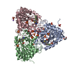 7zsoC 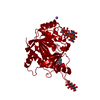 7zspC  7zsqC  7zsrC  8c25C  3phbS S: Starting model for refinement C: citing same article ( |
|---|---|
| Similar structure data |
-Links
 PDBj
PDBj
- Assembly
| Deposited unit | 
| |||||||||||||||||||||||||||||||||||||||||||||||||||||||||||||||||||||||||||||||||||||||||||||||||||||||||||||||||||||||||||||||||||||||||||||||||||||||||||||||||||||||||||||||||||||||||||||||||||||||||||||||||||||||||||||||||||||||
|---|---|---|---|---|---|---|---|---|---|---|---|---|---|---|---|---|---|---|---|---|---|---|---|---|---|---|---|---|---|---|---|---|---|---|---|---|---|---|---|---|---|---|---|---|---|---|---|---|---|---|---|---|---|---|---|---|---|---|---|---|---|---|---|---|---|---|---|---|---|---|---|---|---|---|---|---|---|---|---|---|---|---|---|---|---|---|---|---|---|---|---|---|---|---|---|---|---|---|---|---|---|---|---|---|---|---|---|---|---|---|---|---|---|---|---|---|---|---|---|---|---|---|---|---|---|---|---|---|---|---|---|---|---|---|---|---|---|---|---|---|---|---|---|---|---|---|---|---|---|---|---|---|---|---|---|---|---|---|---|---|---|---|---|---|---|---|---|---|---|---|---|---|---|---|---|---|---|---|---|---|---|---|---|---|---|---|---|---|---|---|---|---|---|---|---|---|---|---|---|---|---|---|---|---|---|---|---|---|---|---|---|---|---|---|---|---|---|---|---|---|---|---|---|---|---|---|---|---|---|---|---|---|
| 1 | 
| |||||||||||||||||||||||||||||||||||||||||||||||||||||||||||||||||||||||||||||||||||||||||||||||||||||||||||||||||||||||||||||||||||||||||||||||||||||||||||||||||||||||||||||||||||||||||||||||||||||||||||||||||||||||||||||||||||||||
| 2 | 
| |||||||||||||||||||||||||||||||||||||||||||||||||||||||||||||||||||||||||||||||||||||||||||||||||||||||||||||||||||||||||||||||||||||||||||||||||||||||||||||||||||||||||||||||||||||||||||||||||||||||||||||||||||||||||||||||||||||||
| Unit cell |
| |||||||||||||||||||||||||||||||||||||||||||||||||||||||||||||||||||||||||||||||||||||||||||||||||||||||||||||||||||||||||||||||||||||||||||||||||||||||||||||||||||||||||||||||||||||||||||||||||||||||||||||||||||||||||||||||||||||||
| Noncrystallographic symmetry (NCS) | NCS domain:
NCS domain segments: Component-ID: _ / Refine code: _
|