 Movie
Movie Controller

[English] 日本語
 Yorodumi
- PDB-7z7q: Structure of the T207D single-point mutant of the fluorescent pro... -
+ Open data

- Basic information
| Entry | Database: PDB / ID: 7z7q | |||||||||
|---|---|---|---|---|---|---|---|---|---|---|
| Title | Structure of the T207D single-point mutant of the fluorescent protein NeonCyan1 at pH 6.5 | |||||||||
 Components Components | NeonCyan1 | |||||||||
 Keywords Keywords | FLUORESCENT PROTEIN / mNeonGreen / Trp-based fluorescent chromophore / rational design | |||||||||
| Function / homology | Green Fluorescent Protein / Green fluorescent protein / Green fluorescent protein-related / Green fluorescent protein / Green fluorescent protein / bioluminescence / Beta Barrel / Mainly Beta / Green fluorescent protein blFP-Y3 Function and homology information Function and homology information | |||||||||
| Biological species |  | |||||||||
| Method |  X-RAY DIFFRACTION / SYNCHROTRON / MOLECULAR REPLACEMENT / Resolution: 1.6 Å X-RAY DIFFRACTION / SYNCHROTRON / MOLECULAR REPLACEMENT / Resolution: 1.6 Å | |||||||||
 Authors Authors | Duarte, K. / Dupuy, J. / Royant, A. | |||||||||
| Funding support |  France, France,  United States, 2items United States, 2items
| |||||||||
 Citation Citation | Journal: Protein Eng.Des.Sel. / Year: 2022 Title: Cyan fluorescent proteins derived from mNeonGreen. Authors: Zarowny, L. / Clavel, D. / Johannson, R. / Duarte, K. / Depernet, H. / Dupuy, J. / Baker, H. / Brown, A. / Royant, A. / Campbell, R.E. | |||||||||
| History |
|
- Structure visualization
| Structure viewer | Molecule: MolmilJmol/JSmol |
|---|
- Downloads & links
-Download
| PDBx/mmCIF format | 7z7q.cif.gz | 190.4 KB | Display | PDBx/mmCIF format |
|---|---|---|---|---|
| PDB format | pdb7z7q.ent.gz | 149 KB | Display | PDB format |
| PDBx/mmJSON format | 7z7q.json.gz | Tree view | PDBx/mmJSON format | |
| Others |  Other downloads Other downloads |
-Validation report
| Summary document | 7z7q_validation.pdf.gz | 470.3 KB | Display | wwPDB validaton report |
|---|---|---|---|---|
| Full document | 7z7q_full_validation.pdf.gz | 480 KB | Display | |
| Data in XML | 7z7q_validation.xml.gz | 36.1 KB | Display | |
| Data in CIF | 7z7q_validation.cif.gz | 50.2 KB | Display | |
| Arichive directory | https://data.pdbj.org/pub/pdb/validation_reports/z7/7z7qftp://data.pdbj.org/pub/pdb/validation_reports/z7/7z7q | HTTPS FTP |
-Related structure data
| Related structure data |  7z7oC  7z7pC 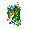 5ltrS S: Starting model for refinement C: citing same article ( |
|---|---|
| Similar structure data |
-Links
 PDBj
PDBj

- Assembly
| Deposited unit | 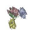
| ||||||||
|---|---|---|---|---|---|---|---|---|---|
| 1 | 
| ||||||||
| 2 | 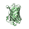
| ||||||||
| 3 | 
| ||||||||
| 4 | 
| ||||||||
| Unit cell |
|
-Components
| #1: Protein | Mass: 26656.734 Da / Num. of mol.: 4 Source method: isolated from a genetically manipulated source Details: Residue CRC69 is formed by the autocatalytic cyclisation of the consecutive amino acid residues GWG, hence the mismatch in the sequence alignment Source: (gene. exp.)  #2: Water | ChemComp-HOH / |  Mass: 18.015 Da / Num. of mol.: 345 / Source method: isolated from a natural source / Formula: H2O Mass: 18.015 Da / Num. of mol.: 345 / Source method: isolated from a natural source / Formula: H2OHas ligand of interest | Y | Has protein modification | Y | |
|---|
-Experimental details
-Experiment
| Experiment | Method: X-RAY DIFFRACTION / Number of used crystals: 1 |
|---|
- Sample preparation
| Crystal grow | Temperature: 293 K / Method: vapor diffusion, hanging drop / pH: 6.5 / Details: 100 mM HEPES pH 6.5 and 22% PEG8000 (w/v) |
|---|
-Data collection
| Diffraction | Mean temperature: 100 K / Serial crystal experiment: N |
|---|---|
| Diffraction source | Source: SYNCHROTRON / Site: ALBA  / Beamline: XALOC / Wavelength: 0.979 Å / Beamline: XALOC / Wavelength: 0.979 Å |
| Detector | Type: DECTRIS PILATUS 6M / Detector: PIXEL / Date: Dec 12, 2019 |
| Radiation | Monochromator: channel-cut Si(111) / Protocol: SINGLE WAVELENGTH / Monochromatic (M) / Laue (L): M / Scattering type: x-ray |
| Radiation wavelength | Wavelength: 0.979 Å / Relative weight: 1 |
| Reflection | Resolution: 1.6→40.06 Å / Num. obs: 85638 / % possible obs: 86.7 % / Redundancy: 1.7 % / Biso Wilson estimate: 16.6 Å2 / CC1/2: 0.993 / Rrim(I) all: 0.109 / Net I/σ(I): 8.8 |
| Reflection shell | Resolution: 1.6→1.66 Å / Redundancy: 2.6 % / Mean I/σ(I) obs: 2.4 / Num. unique obs: 8266 / CC1/2: 0.703 / Rrim(I) all: 0.754 / % possible all: 83.6 |
- Processing
| Software |
| ||||||||||||||||||||||||||||||||||||||||||||||||||||||||||||
|---|---|---|---|---|---|---|---|---|---|---|---|---|---|---|---|---|---|---|---|---|---|---|---|---|---|---|---|---|---|---|---|---|---|---|---|---|---|---|---|---|---|---|---|---|---|---|---|---|---|---|---|---|---|---|---|---|---|---|---|---|---|
| Refinement | Method to determine structure: MOLECULAR REPLACEMENT Starting model: 5LTR Resolution: 1.6→40.06 Å / Cor.coef. Fo:Fc: 0.943 / Cor.coef. Fo:Fc free: 0.932 / SU B: 3.136 / SU ML: 0.104 / Cross valid method: THROUGHOUT / σ(F): 0 / ESU R: 0.143 / ESU R Free: 0.13 / Stereochemistry target values: MAXIMUM LIKELIHOOD Details: HYDROGENS HAVE BEEN ADDED IN THE RIDING POSITIONS U VALUES : REFINED INDIVIDUALLY
| ||||||||||||||||||||||||||||||||||||||||||||||||||||||||||||
| Solvent computation | Ion probe radii: 0.8 Å / Shrinkage radii: 0.8 Å / VDW probe radii: 1.2 Å / Solvent model: MASK | ||||||||||||||||||||||||||||||||||||||||||||||||||||||||||||
| Displacement parameters | Biso max: 52.44 Å2 / Biso mean: 15.588 Å2 / Biso min: 4.1 Å2
| ||||||||||||||||||||||||||||||||||||||||||||||||||||||||||||
| Refinement step | Cycle: final / Resolution: 1.6→40.06 Å
| ||||||||||||||||||||||||||||||||||||||||||||||||||||||||||||
| Refine LS restraints |
| ||||||||||||||||||||||||||||||||||||||||||||||||||||||||||||
| LS refinement shell | Resolution: 1.6→1.64 Å / Rfactor Rfree error: 0
|
