 Movie
Movie Controller

[English] 日本語
 Yorodumi
- PDB-7z7e: Crystal structure of p63 DNA binding domain in complex with inhib... -
+ Open data

- Basic information
| Entry | Database: PDB / ID: 7z7e | ||||||||||||
|---|---|---|---|---|---|---|---|---|---|---|---|---|---|
| Title | Crystal structure of p63 DNA binding domain in complex with inhibitory DARPin G4 | ||||||||||||
 Components Components |
| ||||||||||||
 Keywords Keywords | TRANSCRIPTION / p63 DBD / DARPin / Inhibitor | ||||||||||||
| Function / homology |  Function and homology information Function and homology informationectoderm and mesoderm interaction / epidermal cell division / cloacal septation / positive regulation of somatic stem cell population maintenance / negative regulation of mesoderm development / prostatic bud formation / female genitalia morphogenesis / positive regulation of keratinocyte proliferation / establishment of planar polarity / squamous basal epithelial stem cell differentiation involved in prostate gland acinus development ...ectoderm and mesoderm interaction / epidermal cell division / cloacal septation / positive regulation of somatic stem cell population maintenance / negative regulation of mesoderm development / prostatic bud formation / female genitalia morphogenesis / positive regulation of keratinocyte proliferation / establishment of planar polarity / squamous basal epithelial stem cell differentiation involved in prostate gland acinus development / negative regulation of keratinocyte differentiation / polarized epithelial cell differentiation / proximal/distal pattern formation / positive regulation of fibroblast apoptotic process / skin morphogenesis / positive regulation of cell cycle G1/S phase transition / negative regulation of intracellular estrogen receptor signaling pathway / sympathetic nervous system development / cranial skeletal system development / post-anal tail morphogenesis / embryonic forelimb morphogenesis / Differentiation of Keratinocytes in Interfollicular Epidermis in Mammalian Skin / embryonic hindlimb morphogenesis / TP53 Regulates Transcription of Death Receptors and Ligands / Activation of PUMA and translocation to mitochondria / Regulation of TP53 Activity through Association with Co-factors / hair follicle morphogenesis / WW domain binding / epithelial cell development / TP53 Regulates Transcription of Caspase Activators and Caspases / positive regulation of Notch signaling pathway / regulation of epidermal cell division / positive regulation of stem cell proliferation / odontogenesis of dentin-containing tooth / TP53 Regulates Transcription of Genes Involved in Cytochrome C Release / TP53 regulates transcription of several additional cell death genes whose specific roles in p53-dependent apoptosis remain uncertain / negative regulation of cellular senescence / keratinocyte proliferation / intrinsic apoptotic signaling pathway in response to DNA damage by p53 class mediator / Pyroptosis / establishment of skin barrier / positive regulation of osteoblast differentiation / keratinocyte differentiation / Notch signaling pathway / MDM2/MDM4 family protein binding / positive regulation of apoptotic signaling pathway / stem cell proliferation / skeletal system development / determination of adult lifespan / TP53 Regulates Metabolic Genes / RNA polymerase II transcription regulatory region sequence-specific DNA binding / protein tetramerization / promoter-specific chromatin binding / p53 binding / cellular senescence / neuron apoptotic process / DNA-binding transcription activator activity, RNA polymerase II-specific / spermatogenesis / damaged DNA binding / transcription by RNA polymerase II / DNA-binding transcription factor activity, RNA polymerase II-specific / RNA polymerase II cis-regulatory region sequence-specific DNA binding / chromatin remodeling / DNA-binding transcription factor activity / negative regulation of DNA-templated transcription / apoptotic process / dendrite / DNA damage response / chromatin binding / regulation of transcription by RNA polymerase II / chromatin / positive regulation of DNA-templated transcription / negative regulation of transcription by RNA polymerase II / positive regulation of transcription by RNA polymerase II / protein-containing complex / DNA binding / nucleoplasm / metal ion binding / identical protein binding / nucleus / cytoplasm Similarity search - Function | ||||||||||||
| Biological species |  Homo sapiens (human) Homo sapiens (human)synthetic construct (others) | ||||||||||||
| Method |  X-RAY DIFFRACTION / SYNCHROTRON / MOLECULAR REPLACEMENT / Resolution: 1.8 Å X-RAY DIFFRACTION / SYNCHROTRON / MOLECULAR REPLACEMENT / Resolution: 1.8 Å | ||||||||||||
 Authors Authors | Strubel, A. / Gebel, J. / Chaikuad, A. / Muenick, P. / Doetsch, V. | ||||||||||||
| Funding support |  Germany, Germany,  Canada, 3items Canada, 3items
| ||||||||||||
 Citation Citation | Journal: Cell Death Differ. / Year: 2022 Title: Designed Ankyrin Repeat Proteins as a tool box for analyzing p63. Authors: Strubel, A. / Munick, P. / Chaikuad, A. / Dreier, B. / Schaefer, J. / Gebel, J. / Osterburg, C. / Tuppi, M. / Schafer, B. / Knapp, S. / Pluckthun, A. / Dotsch, V. | ||||||||||||
| History |
|
- Structure visualization
| Structure viewer | Molecule: MolmilJmol/JSmol |
|---|
- Downloads & links
-Download
| PDBx/mmCIF format | 7z7e.cif.gz | 148.8 KB | Display | PDBx/mmCIF format |
|---|---|---|---|---|
| PDB format | pdb7z7e.ent.gz | 111.5 KB | Display | PDB format |
| PDBx/mmJSON format | 7z7e.json.gz | Tree view | PDBx/mmJSON format | |
| Others |  Other downloads Other downloads |
-Validation report
| Summary document | 7z7e_validation.pdf.gz | 615.9 KB | Display | wwPDB validaton report |
|---|---|---|---|---|
| Full document | 7z7e_full_validation.pdf.gz | 617.4 KB | Display | |
| Data in XML | 7z7e_validation.xml.gz | 15.9 KB | Display | |
| Data in CIF | 7z7e_validation.cif.gz | 23 KB | Display | |
| Arichive directory | https://data.pdbj.org/pub/pdb/validation_reports/z7/7z7eftp://data.pdbj.org/pub/pdb/validation_reports/z7/7z7e | HTTPS FTP |
-Related structure data
| Related structure data | 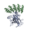 7z71C 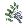 7z72C 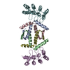 7z73C 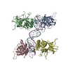 3us0S S: Starting model for refinement C: citing same article ( |
|---|---|
| Similar structure data |
-Links
 PDBj
PDBj


- Assembly
| Deposited unit | 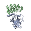
| ||||||||
|---|---|---|---|---|---|---|---|---|---|
| 1 |
| ||||||||
| Unit cell |
|
-Components
| #1: Protein | Mass: 22752.920 Da / Num. of mol.: 1 Source method: isolated from a genetically manipulated source Source: (gene. exp.) Homo sapiens (human) / Gene: TP63, KET, P63, P73H, P73L, TP73L / Production host:  |
|---|---|
| #2: Protein | Mass: 16908.893 Da / Num. of mol.: 1 Source method: isolated from a genetically manipulated source Source: (gene. exp.) synthetic construct (others) / Production host: |
| #3: Chemical | ChemComp-ZN /   Mass: 65.409 Da / Num. of mol.: 1 / Source method: obtained synthetically / Formula: Zn / Feature type: SUBJECT OF INVESTIGATION Mass: 65.409 Da / Num. of mol.: 1 / Source method: obtained synthetically / Formula: Zn / Feature type: SUBJECT OF INVESTIGATION |
| #4: Water | ChemComp-HOH /  Mass: 18.015 Da / Num. of mol.: 186 / Source method: isolated from a natural source / Formula: H2O Mass: 18.015 Da / Num. of mol.: 186 / Source method: isolated from a natural source / Formula: H2O |
| Has ligand of interest | Y |
-Experimental details
-Experiment
| Experiment | Method: X-RAY DIFFRACTION / Number of used crystals: 1 |
|---|
- Sample preparation
| Crystal | Density Matthews: 2.62 Å3/Da / Density % sol: 52.98 % |
|---|---|
| Crystal grow | Temperature: 295 K / Method: vapor diffusion / pH: 7.5 / Details: 25% PEG 3350 0.2M Li2SO4 0.1M HEPES / PH range: 7.5 |
-Data collection
| Diffraction | Mean temperature: 100 K / Serial crystal experiment: N | |||||||||||||||||||||
|---|---|---|---|---|---|---|---|---|---|---|---|---|---|---|---|---|---|---|---|---|---|---|
| Diffraction source | Source: SYNCHROTRON / Site: SLS  / Beamline: X06SA / Wavelength: 1 Å / Beamline: X06SA / Wavelength: 1 Å | |||||||||||||||||||||
| Detector | Type: DECTRIS EIGER X 16M / Detector: PIXEL / Date: Dec 15, 2021 | |||||||||||||||||||||
| Radiation | Protocol: SINGLE WAVELENGTH / Monochromatic (M) / Laue (L): M / Scattering type: x-ray | |||||||||||||||||||||
| Radiation wavelength | Wavelength: 1 Å / Relative weight: 1 | |||||||||||||||||||||
| Reflection | Resolution: 1.8→48.29 Å / Num. obs: 37301 / % possible obs: 96.4 % / Redundancy: 1.9 % / CC1/2: 0.999 / Rmerge(I) obs: 0.012 / Rpim(I) all: 0.019 / Rrim(I) all: 0.027 / Net I/σ(I): 23 | |||||||||||||||||||||
| Reflection shell | Diffraction-ID: 1
|
- Processing
| Software |
| ||||||||||||||||||||||||||||||||||||||||||||||||||||||||||||||||||||||||||||||||||||||||||||||||||||||||||||||||||||||||||||||||||||||||||||||||||||||
|---|---|---|---|---|---|---|---|---|---|---|---|---|---|---|---|---|---|---|---|---|---|---|---|---|---|---|---|---|---|---|---|---|---|---|---|---|---|---|---|---|---|---|---|---|---|---|---|---|---|---|---|---|---|---|---|---|---|---|---|---|---|---|---|---|---|---|---|---|---|---|---|---|---|---|---|---|---|---|---|---|---|---|---|---|---|---|---|---|---|---|---|---|---|---|---|---|---|---|---|---|---|---|---|---|---|---|---|---|---|---|---|---|---|---|---|---|---|---|---|---|---|---|---|---|---|---|---|---|---|---|---|---|---|---|---|---|---|---|---|---|---|---|---|---|---|---|---|---|---|---|---|
| Refinement | Method to determine structure: MOLECULAR REPLACEMENT Starting model: 3US0 Resolution: 1.8→48.29 Å / Cor.coef. Fo:Fc: 0.967 / Cor.coef. Fo:Fc free: 0.946 / SU B: 3.33 / SU ML: 0.1 / Cross valid method: FREE R-VALUE / ESU R: 0.126 / ESU R Free: 0.131 Details: Hydrogens have been added in their riding positions
| ||||||||||||||||||||||||||||||||||||||||||||||||||||||||||||||||||||||||||||||||||||||||||||||||||||||||||||||||||||||||||||||||||||||||||||||||||||||
| Solvent computation | Ion probe radii: 0.8 Å / Shrinkage radii: 0.8 Å / VDW probe radii: 1.2 Å / Solvent model: MASK BULK SOLVENT | ||||||||||||||||||||||||||||||||||||||||||||||||||||||||||||||||||||||||||||||||||||||||||||||||||||||||||||||||||||||||||||||||||||||||||||||||||||||
| Displacement parameters | Biso mean: 40.609 Å2
| ||||||||||||||||||||||||||||||||||||||||||||||||||||||||||||||||||||||||||||||||||||||||||||||||||||||||||||||||||||||||||||||||||||||||||||||||||||||
| Refinement step | Cycle: LAST / Resolution: 1.8→48.29 Å
| ||||||||||||||||||||||||||||||||||||||||||||||||||||||||||||||||||||||||||||||||||||||||||||||||||||||||||||||||||||||||||||||||||||||||||||||||||||||
| Refine LS restraints |
| ||||||||||||||||||||||||||||||||||||||||||||||||||||||||||||||||||||||||||||||||||||||||||||||||||||||||||||||||||||||||||||||||||||||||||||||||||||||
| LS refinement shell |
|
