 Movie
Movie Controller

[English] 日本語
 Yorodumi
- PDB-7yzi: Structure of Mycobacterium tuberculosis adenylyl cyclase Rv1625c / Cya -
+ Open data

- Basic information
| Entry | Database: PDB / ID: 7yzi | |||||||||||||||||||||||||||||||||||||||
|---|---|---|---|---|---|---|---|---|---|---|---|---|---|---|---|---|---|---|---|---|---|---|---|---|---|---|---|---|---|---|---|---|---|---|---|---|---|---|---|---|
| Title | Structure of Mycobacterium tuberculosis adenylyl cyclase Rv1625c / Cya | |||||||||||||||||||||||||||||||||||||||
 Components Components |
| |||||||||||||||||||||||||||||||||||||||
 Keywords Keywords | MEMBRANE PROTEIN / Adenylyl cyclase / cyclic adenosine monophosphate / signal transduction / nanobody | |||||||||||||||||||||||||||||||||||||||
| Function / homology |  Function and homology information Function and homology informationreceptor guanylyl cyclase signaling pathway / peptide receptor activity / cGMP biosynthetic process / guanylate cyclase activity / adenylate cyclase / adenylate cyclase activity / cAMP biosynthetic process / manganese ion binding / intracellular signal transduction / magnesium ion binding ...receptor guanylyl cyclase signaling pathway / peptide receptor activity / cGMP biosynthetic process / guanylate cyclase activity / adenylate cyclase / adenylate cyclase activity / cAMP biosynthetic process / manganese ion binding / intracellular signal transduction / magnesium ion binding / ATP binding / plasma membrane Similarity search - Function | |||||||||||||||||||||||||||||||||||||||
| Biological species |  Mycobacterium tuberculosis '98-R604 INH-RIF-EM' (bacteria) Mycobacterium tuberculosis '98-R604 INH-RIF-EM' (bacteria) | |||||||||||||||||||||||||||||||||||||||
| Method | ELECTRON MICROSCOPY / single particle reconstruction / cryo EM / Resolution: 3.83 Å | |||||||||||||||||||||||||||||||||||||||
 Authors Authors | Mehta, V. / Khanppnavar, B. / Korkhov, V.M. | |||||||||||||||||||||||||||||||||||||||
| Funding support |  Switzerland, 3items Switzerland, 3items
| |||||||||||||||||||||||||||||||||||||||
 Citation Citation | Journal: Elife / Year: 2022 Title: Structure of Cya, an evolutionary ancestor of the mammalian membrane adenylyl cyclases. Authors: Ved Mehta / Basavraj Khanppnavar / Dina Schuster / Ilayda Kantarci / Irene Vercellino / Angela Kosturanova / Tarun Iype / Sasa Stefanic / Paola Picotti / Volodymyr M Korkhov / Abstract: adenylyl cyclase (AC) Rv1625c/Cya is an evolutionary ancestor of the mammalian membrane ACs and a model system for studies of their structure and function. Although the vital role of ACs in cellular ... adenylyl cyclase (AC) Rv1625c/Cya is an evolutionary ancestor of the mammalian membrane ACs and a model system for studies of their structure and function. Although the vital role of ACs in cellular signalling is well established, the function of their transmembrane (TM) regions remains unknown. Here, we describe the cryo-EM structure of Cya bound to a stabilizing nanobody at 3.6 Å resolution. The TM helices 1-5 form a structurally conserved domain that facilitates the assembly of the helical and catalytic domains. The TM region contains discrete pockets accessible from the extracellular and cytosolic side of the membrane. Neutralization of the negatively charged extracellular pocket Ex1 destabilizes the cytosolic helical domain and reduces the catalytic activity of the enzyme. The TM domain acts as a functional component of Cya, guiding the assembly of the catalytic domain and providing the means for direct regulation of catalytic activity in response to extracellular ligands. | |||||||||||||||||||||||||||||||||||||||
| History |
|
- Structure visualization
| Structure viewer | Molecule: MolmilJmol/JSmol |
|---|
- Downloads & links
-Download
| PDBx/mmCIF format | 7yzi.cif.gz | 371.5 KB | Display | PDBx/mmCIF format |
|---|---|---|---|---|
| PDB format | pdb7yzi.ent.gz | 302.6 KB | Display | PDB format |
| PDBx/mmJSON format | 7yzi.json.gz | Tree view | PDBx/mmJSON format | |
| Others |  Other downloads Other downloads |
-Validation report
| Arichive directory | https://data.pdbj.org/pub/pdb/validation_reports/yz/7yziftp://data.pdbj.org/pub/pdb/validation_reports/yz/7yzi | HTTPS FTP |
|---|
-Related structure data
| Related structure data |  14388MC 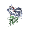 7yz9C  7yzkC M: map data used to model this data C: citing same article ( |
|---|---|
| Similar structure data |

-Links
 PDBj
PDBj


- Assembly
| Deposited unit | 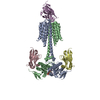
|
|---|---|
| 1 |
|
-Components
| #1: Protein | Mass: 50386.613 Da / Num. of mol.: 2 Source method: isolated from a genetically manipulated source Source: (gene. exp.) Mycobacterium tuberculosis '98-R604 INH-RIF-EM' (bacteria)Gene: cya, Rv1625c, MTCY01B2.17c / Production host: #2: Antibody | Mass: 13771.179 Da / Num. of mol.: 3 Source method: isolated from a genetically manipulated source Source: (gene. exp.) #3: Chemical |   Mass: 656.328 Da / Num. of mol.: 2 / Source method: obtained synthetically / Formula: C18H23N6O15P3 Mass: 656.328 Da / Num. of mol.: 2 / Source method: obtained synthetically / Formula: C18H23N6O15P3#4: Chemical | ChemComp-MN /   Mass: 54.938 Da / Num. of mol.: 4 / Source method: obtained synthetically / Formula: Mn Mass: 54.938 Da / Num. of mol.: 4 / Source method: obtained synthetically / Formula: MnHas ligand of interest | N | Has protein modification | Y | |
|---|
-Experimental details
-Experiment
| Experiment | Method: ELECTRON MICROSCOPY |
|---|---|
| EM experiment | Aggregation state: PARTICLE / 3D reconstruction method: single particle reconstruction |
- Sample preparation
| Component |
| ||||||||||||||||||||||||
|---|---|---|---|---|---|---|---|---|---|---|---|---|---|---|---|---|---|---|---|---|---|---|---|---|---|
| Molecular weight | Experimental value: NO | ||||||||||||||||||||||||
| Source (natural) |
| ||||||||||||||||||||||||
| Source (recombinant) |
| ||||||||||||||||||||||||
| Buffer solution | pH: 7.5 / Details: 50 mM Tris pH 7.5, 200 mM NaCl, 0.1 % digitonin | ||||||||||||||||||||||||
| Specimen | Embedding applied: NO / Shadowing applied: NO / Staining applied: NO / Vitrification applied: YES | ||||||||||||||||||||||||
| Vitrification | Cryogen name: ETHANE |
- Electron microscopy imaging
| Experimental equipment |  Model: Titan Krios / Image courtesy: FEI Company |
|---|---|
| Microscopy | Model: TFS KRIOS |
| Electron gun | Electron source:  FIELD EMISSION GUN / Accelerating voltage: 300 kV / Illumination mode: FLOOD BEAM FIELD EMISSION GUN / Accelerating voltage: 300 kV / Illumination mode: FLOOD BEAM |
| Electron lens | Mode: BRIGHT FIELD / Nominal defocus max: 3000 nm / Nominal defocus min: 500 nm |
| Image recording | Electron dose: 48 e/Å2 / Film or detector model: GATAN K3 (6k x 4k) |
- Processing
| Software | Name: PHENIX / Version: 1.19.2_4158: / Classification: refinement | ||||||||||||||||||||||||||||
|---|---|---|---|---|---|---|---|---|---|---|---|---|---|---|---|---|---|---|---|---|---|---|---|---|---|---|---|---|---|
| EM software |
| ||||||||||||||||||||||||||||
| CTF correction | Type: NONE | ||||||||||||||||||||||||||||
| Symmetry | Point symmetry: C1 (asymmetric) | ||||||||||||||||||||||||||||
| 3D reconstruction | Resolution: 3.83 Å / Resolution method: FSC 0.143 CUT-OFF / Num. of particles: 646042 / Symmetry type: POINT | ||||||||||||||||||||||||||||
| Refine LS restraints |
|
