+ Open data
- Basic information
| Entry | Database: PDB / ID: 7yz4 | |||||||||
|---|---|---|---|---|---|---|---|---|---|---|
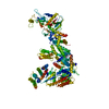
| Title | Mouse endoribonuclease Dicer (composite structure) | |||||||||
 Components Components | Endoribonuclease Dicer | |||||||||
 Keywords Keywords | RNA BINDING PROTEIN / endoribonuclease / gene silencing / post-transcriptional / catalytic enzyme / cytoplasm | |||||||||
| Function / homology |  Function and homology information Function and homology informationregulation of muscle cell apoptotic process / MicroRNA (miRNA) biogenesis / Small interfering RNA (siRNA) biogenesis / ganglion development / hair follicle cell proliferation / zygote asymmetric cell division / cardiac neural crest cell development involved in outflow tract morphogenesis / regulation of oligodendrocyte differentiation / olfactory bulb interneuron differentiation / regulation of odontogenesis of dentin-containing tooth ...regulation of muscle cell apoptotic process / MicroRNA (miRNA) biogenesis / Small interfering RNA (siRNA) biogenesis / ganglion development / hair follicle cell proliferation / zygote asymmetric cell division / cardiac neural crest cell development involved in outflow tract morphogenesis / regulation of oligodendrocyte differentiation / olfactory bulb interneuron differentiation / regulation of odontogenesis of dentin-containing tooth / regulation of enamel mineralization / regulation of miRNA metabolic process / peripheral nervous system myelin formation / trophectodermal cell proliferation / spermatogonial cell division / regulation of RNA metabolic process / regulation of epithelial cell differentiation / global gene silencing by mRNA cleavage / spinal cord motor neuron differentiation / negative regulation of Schwann cell proliferation / epidermis morphogenesis / reproductive structure development / ribonuclease III / myoblast differentiation involved in skeletal muscle regeneration / regulation of Notch signaling pathway / regulation of regulatory T cell differentiation / positive regulation of Schwann cell differentiation / nerve development / RISC-loading complex / positive regulation of myelination / meiotic spindle organization / intestinal epithelial cell development / RISC complex assembly / miRNA processing / regulatory ncRNA-mediated post-transcriptional gene silencing / ribonuclease III activity / pre-miRNA processing / siRNA processing / pericentric heterochromatin formation / regulation of stem cell differentiation / regulation of viral genome replication / RISC complex / mRNA stabilization / inner ear receptor cell development / cartilage development / embryonic limb morphogenesis / embryonic hindlimb morphogenesis / digestive tract development / positive regulation of miRNA metabolic process / cardiac muscle cell development / regulation of neuron differentiation / miRNA binding / regulation of myelination / negative regulation of glial cell proliferation / hair follicle morphogenesis / stem cell population maintenance / branching morphogenesis of an epithelial tube / hair follicle development / regulation of neurogenesis / RNA processing / postsynaptic density, intracellular component / spleen development / neuron projection morphogenesis / spindle assembly / lung development / post-embryonic development / helicase activity / cerebral cortex development / multicellular organism growth / rRNA processing / regulation of gene expression / regulation of inflammatory response / angiogenesis / gene expression / defense response to virus / cell population proliferation / regulation of cell cycle / positive regulation of gene expression / perinuclear region of cytoplasm / glutamatergic synapse / negative regulation of transcription by RNA polymerase II / positive regulation of transcription by RNA polymerase II / DNA binding / ATP binding / metal ion binding / cytosol / cytoplasm Similarity search - Function | |||||||||
| Biological species |  | |||||||||
| Method | ELECTRON MICROSCOPY / single particle reconstruction / cryo EM / Resolution: 3.84 Å | |||||||||
 Authors Authors | Zanova, M. / Zapletal, D. / Kubicek, K. / Stefl, R. / Pinkas, M. / Novacek, J. | |||||||||
| Funding support |  Czech Republic, 2items Czech Republic, 2items
| |||||||||
 Citation Citation |  Journal: Mol Cell / Year: 2022 Journal: Mol Cell / Year: 2022Title: Structural and functional basis of mammalian microRNA biogenesis by Dicer. Authors: David Zapletal / Eliska Taborska / Josef Pasulka / Radek Malik / Karel Kubicek / Martina Zanova / Christian Much / Marek Sebesta / Valeria Buccheri / Filip Horvat / Irena Jenickova / ...Authors: David Zapletal / Eliska Taborska / Josef Pasulka / Radek Malik / Karel Kubicek / Martina Zanova / Christian Much / Marek Sebesta / Valeria Buccheri / Filip Horvat / Irena Jenickova / Michaela Prochazkova / Jan Prochazka / Matyas Pinkas / Jiri Novacek / Diego F Joseph / Radislav Sedlacek / Carrie Bernecky / Dónal O'Carroll / Richard Stefl / Petr Svoboda /      Abstract: MicroRNA (miRNA) and RNA interference (RNAi) pathways rely on small RNAs produced by Dicer endonucleases. Mammalian Dicer primarily supports the essential gene-regulating miRNA pathway, but how it is ...MicroRNA (miRNA) and RNA interference (RNAi) pathways rely on small RNAs produced by Dicer endonucleases. Mammalian Dicer primarily supports the essential gene-regulating miRNA pathway, but how it is specifically adapted to miRNA biogenesis is unknown. We show that the adaptation entails a unique structural role of Dicer's DExD/H helicase domain. Although mice tolerate loss of its putative ATPase function, the complete absence of the domain is lethal because it assures high-fidelity miRNA biogenesis. Structures of murine Dicer•-miRNA precursor complexes revealed that the DExD/H domain has a helicase-unrelated structural function. It locks Dicer in a closed state, which facilitates miRNA precursor selection. Transition to a cleavage-competent open state is stimulated by Dicer-binding protein TARBP2. Absence of the DExD/H domain or its mutations unlocks the closed state, reduces substrate selectivity, and activates RNAi. Thus, the DExD/H domain structurally contributes to mammalian miRNA biogenesis and underlies mechanistical partitioning of miRNA and RNAi pathways. | |||||||||
| History |
|
- Structure visualization
| Structure viewer | Molecule:  Molmil Molmil Jmol/JSmol Jmol/JSmol |
|---|
- Downloads & links
- Download
| PDBx/mmCIF format |  7yz4.cif.gz 7yz4.cif.gz | 438.4 KB | Display |  PDBx/mmCIF format PDBx/mmCIF format |
|---|---|---|---|---|
| PDB format |  pdb7yz4.ent.gz pdb7yz4.ent.gz | 346.7 KB | Display |  PDB format PDB format |
| PDBx/mmJSON format |  7yz4.json.gz 7yz4.json.gz | Tree view |  PDBx/mmJSON format PDBx/mmJSON format | |
| Others |  Other downloads Other downloads |
-Validation report
| Summary document |  7yz4_validation.pdf.gz 7yz4_validation.pdf.gz | 1.3 MB | Display |  wwPDB validaton report wwPDB validaton report |
|---|---|---|---|---|
| Full document |  7yz4_full_validation.pdf.gz 7yz4_full_validation.pdf.gz | 1.3 MB | Display | |
| Data in XML |  7yz4_validation.xml.gz 7yz4_validation.xml.gz | 49.8 KB | Display | |
| Data in CIF |  7yz4_validation.cif.gz 7yz4_validation.cif.gz | 74.4 KB | Display | |
| Arichive directory |  https://data.pdbj.org/pub/pdb/validation_reports/yz/7yz4 https://data.pdbj.org/pub/pdb/validation_reports/yz/7yz4 ftp://data.pdbj.org/pub/pdb/validation_reports/yz/7yz4 ftp://data.pdbj.org/pub/pdb/validation_reports/yz/7yz4 | HTTPS FTP |
-Related structure data
| Related structure data |  14387MC  7yymC  7yynC  7zpiC  7zpjC  7zpkC C: citing same article ( M: map data used to model this data |
|---|---|
| Similar structure data | Similarity search - Function & homology  F&H Search F&H Search |
- Links
Links
- Assembly
| Deposited unit | 
|
|---|---|
| 1 |
|
- Components
| #1: Protein | Mass: 226809.594 Da / Num. of mol.: 1 Source method: isolated from a genetically manipulated source Source: (gene. exp.)   Baculovirus expression vector pFastBac1-HM / References: UniProt: Q8R418, ribonuclease III Baculovirus expression vector pFastBac1-HM / References: UniProt: Q8R418, ribonuclease III |
|---|
-Experimental details
-Experiment
| Experiment | Method: ELECTRON MICROSCOPY |
|---|---|
| EM experiment | Aggregation state: PARTICLE / 3D reconstruction method: single particle reconstruction |
- Sample preparation
| Component | Name: Free form of mouse somatic dicer / Type: ORGANELLE OR CELLULAR COMPONENT / Entity ID: all / Source: RECOMBINANT | ||||||||||||||||||||
|---|---|---|---|---|---|---|---|---|---|---|---|---|---|---|---|---|---|---|---|---|---|
| Molecular weight | Experimental value: NO | ||||||||||||||||||||
| Source (natural) | Organism:  | ||||||||||||||||||||
| Source (recombinant) | Organism:  Baculovirus expression vector pFastBac1-HM Baculovirus expression vector pFastBac1-HM | ||||||||||||||||||||
| Buffer solution | pH: 8 / Details: The buffer was always prepared fresh | ||||||||||||||||||||
| Buffer component |
| ||||||||||||||||||||
| Specimen | Conc.: 0.2 mg/ml / Embedding applied: NO / Shadowing applied: NO / Staining applied: NO / Vitrification applied: YES | ||||||||||||||||||||
| Vitrification | Instrument: FEI VITROBOT MARK IV / Cryogen name: ETHANE / Humidity: 100 % / Chamber temperature: 277.15 K / Details: Described in STAR methods |
- Electron microscopy imaging
| Experimental equipment |  Model: Titan Krios / Image courtesy: FEI Company |
|---|---|
| Microscopy | Model: FEI TITAN KRIOS |
| Electron gun | Electron source:  FIELD EMISSION GUN / Accelerating voltage: 300 kV / Illumination mode: FLOOD BEAM FIELD EMISSION GUN / Accelerating voltage: 300 kV / Illumination mode: FLOOD BEAM |
| Electron lens | Mode: BRIGHT FIELD / Nominal defocus max: 3500 nm / Nominal defocus min: 800 nm |
| Image recording | Electron dose: 55 e/Å2 / Detector mode: COUNTING / Film or detector model: GATAN K2 SUMMIT (4k x 4k) / Num. of real images: 6354 |
- Processing
| Software | Name: PHENIX / Version: 1.19.2_4158: / Classification: refinement | ||||||||||||||||||||||||||||||||||||||||||||||||
|---|---|---|---|---|---|---|---|---|---|---|---|---|---|---|---|---|---|---|---|---|---|---|---|---|---|---|---|---|---|---|---|---|---|---|---|---|---|---|---|---|---|---|---|---|---|---|---|---|---|
| EM software |
| ||||||||||||||||||||||||||||||||||||||||||||||||
| CTF correction | Type: PHASE FLIPPING AND AMPLITUDE CORRECTION | ||||||||||||||||||||||||||||||||||||||||||||||||
| Particle selection | Num. of particles selected: 862842 | ||||||||||||||||||||||||||||||||||||||||||||||||
| 3D reconstruction | Resolution: 3.84 Å / Resolution method: FSC 0.143 CUT-OFF / Num. of particles: 92906 / Algorithm: FOURIER SPACE Details: The map resolution was further improved up to 3.47 with local refinement of the individual protein domains. Num. of class averages: 81 / Symmetry type: POINT | ||||||||||||||||||||||||||||||||||||||||||||||||
| Atomic model building |
| ||||||||||||||||||||||||||||||||||||||||||||||||
| Refine LS restraints |
|
 Movie
Movie Controller


 PDBj
PDBj

