 Movie
Movie Controller


+ Open data

- Basic information
| Entry |  | |||||||||
|---|---|---|---|---|---|---|---|---|---|---|
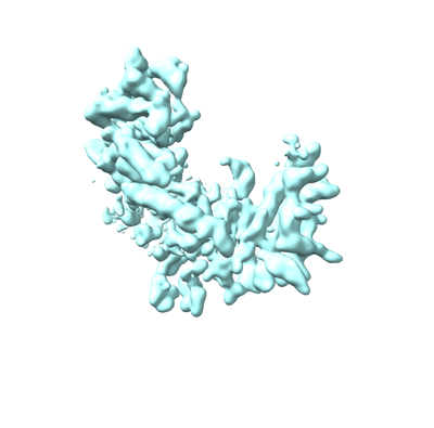
| Title | Mouse endoribonuclease Dicer | |||||||||
 Map data Map data | Locally refined electron density map of Dicer helicase domain | |||||||||
 Sample Sample |
| |||||||||
 Keywords Keywords | endoribonuclease / gene silencing / post-transcriptional / catalytic enzyme / cytoplasm / RNA BINDING PROTEIN | |||||||||
| Biological species |  | |||||||||
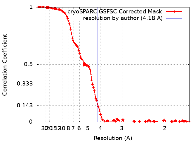
| Method | single particle reconstruction / cryo EM / Resolution: 4.18 Å | |||||||||
 Authors Authors | Zanova M / Zapletal D / Kubicek K / Stefl R / Pinkas M / Novacek J | |||||||||
| Funding support |  Czech Republic, 2 items Czech Republic, 2 items
| |||||||||
 Citation Citation | Journal: To Be Published Title: Mammalian Dicer uses its helicase domain to control RNA processing Authors: Zapletal D / Kubicek K / Zanova M / Sebesta M / Pinkas M / Malik R / Joseph DF / Novacek J / Svoboda P / Stefl R | |||||||||
| History |
|
- Structure visualization
| Supplemental images |
|---|
- Downloads & links
-EMDB archive
| Map data | emd_15299.map.gz | 200.9 MB |  EMDB map data format EMDB map data format | |
|---|---|---|---|---|
| Header (meta data) | emd-15299-v30.xmlemd-15299.xml | 16.1 KB 16.1 KB | Display Display | EMDB header |
| FSC (resolution estimation) | emd_15299_fsc.xml | 12.8 KB | Display | FSC data file |
| Images |  emd_15299.png emd_15299.png | 60.4 KB | ||
| Others | emd_15299_half_map_1.map.gzemd_15299_half_map_2.map.gz | 177.1 MB 177.2 MB | ||
| Archive directory |  http://ftp.pdbj.org/pub/emdb/structures/EMD-15299ftp://ftp.pdbj.org/pub/emdb/structures/EMD-15299 http://ftp.pdbj.org/pub/emdb/structures/EMD-15299ftp://ftp.pdbj.org/pub/emdb/structures/EMD-15299 | HTTPS FTP |
-Related structure data


-Links
| EMDB pages | EMDB (EBI/PDBe) / EMDataResource |
|---|
-Map
| File | Download / File: emd_15299.map.gz / Format: CCP4 / Size: 216 MB / Type: IMAGE STORED AS FLOATING POINT NUMBER (4 BYTES) | ||||||||||||||||||||||||||||||||||||
|---|---|---|---|---|---|---|---|---|---|---|---|---|---|---|---|---|---|---|---|---|---|---|---|---|---|---|---|---|---|---|---|---|---|---|---|---|---|
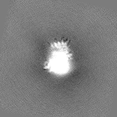
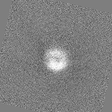
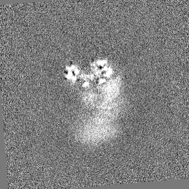
| Annotation | Locally refined electron density map of Dicer helicase domain | ||||||||||||||||||||||||||||||||||||
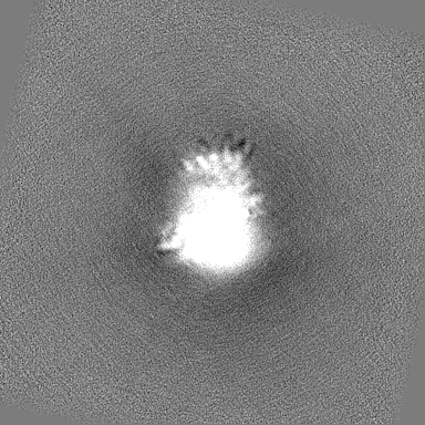
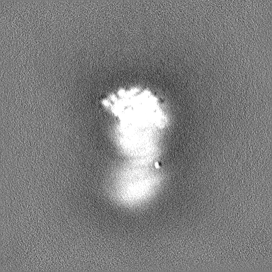
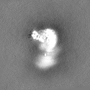
| Projections & slices | Image control
Images are generated by Spider. | ||||||||||||||||||||||||||||||||||||
| Voxel size | X=Y=Z: 0.828 Å | ||||||||||||||||||||||||||||||||||||
| Density |
| ||||||||||||||||||||||||||||||||||||
| Symmetry | Space group: 1 | ||||||||||||||||||||||||||||||||||||
| Details | EMDB XML:
|
 Z (Sec.)
 Y (Row.)
 X (Col.)


-Supplemental data
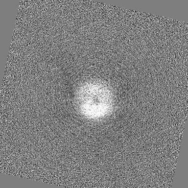
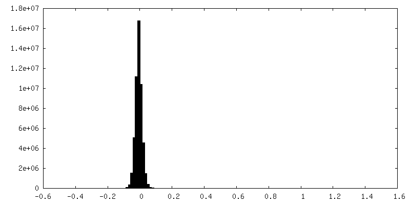
-Half map: half map A
| File | emd_15299_half_map_1.map | ||||||||||||
|---|---|---|---|---|---|---|---|---|---|---|---|---|---|
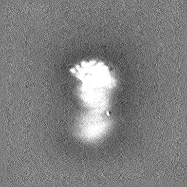
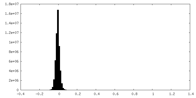
| Annotation | half map A | ||||||||||||
| Projections & Slices |
| ||||||||||||
| Density Histograms |
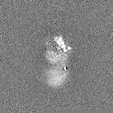

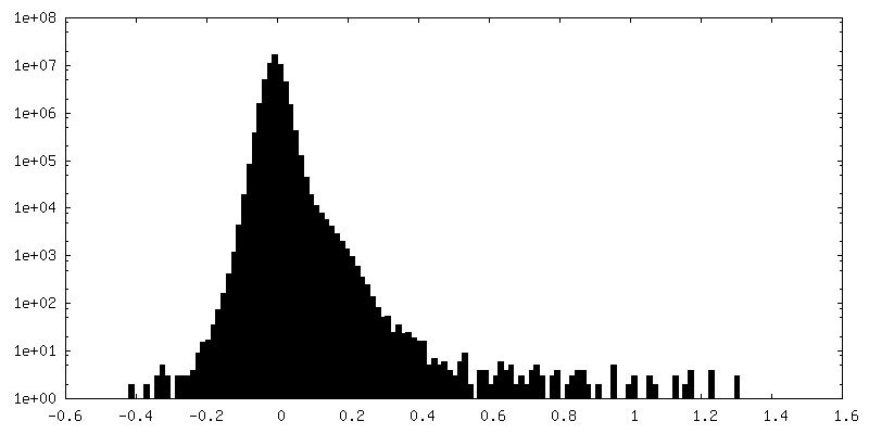
-Half map: half map B
| File | emd_15299_half_map_2.map | ||||||||||||
|---|---|---|---|---|---|---|---|---|---|---|---|---|---|
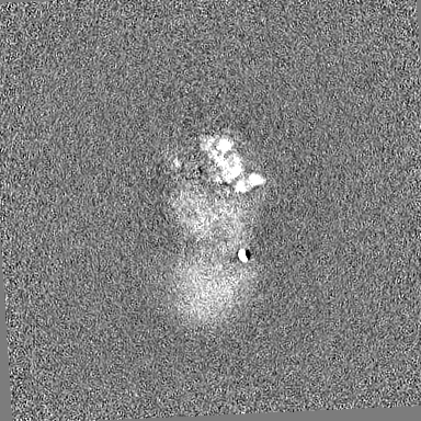
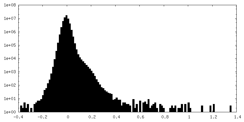
| Annotation | half map B | ||||||||||||
| Projections & Slices |
| ||||||||||||
| Density Histograms |

- Sample components
-Entire : Free form of mouse somatic dicer
| Entire | Name: Free form of mouse somatic dicer |
|---|---|
| Components |
|
-Supramolecule #1: Free form of mouse somatic dicer
| Supramolecule | Name: Free form of mouse somatic dicer / type: organelle_or_cellular_component / ID: 1 / Parent: 0 / Macromolecule list: #1 |
|---|---|
| Source (natural) | Organism: |
-Experimental details
-Structure determination
| Method | cryo EM |
|---|---|
 Processing Processing | single particle reconstruction |
| Aggregation state | particle |
-Sample preparation
| Concentration | 0.20 mg/mL | ||||||||||
|---|---|---|---|---|---|---|---|---|---|---|---|
| Buffer | pH: 8 Component:
Details: The buffer was always prepared fresh | ||||||||||
| Vitrification | Cryogen name: ETHANE / Chamber humidity: 100 % / Chamber temperature: 277.15 K / Instrument: FEI VITROBOT MARK IV / Details: Described in STAR methods. |
- Electron microscopy
| Microscope | FEI TITAN KRIOS |
|---|---|
| Image recording | Film or detector model: GATAN K2 SUMMIT (4k x 4k) / Detector mode: COUNTING / Number real images: 6354 / Average electron dose: 55.0 e/Å2 |
| Electron beam | Acceleration voltage: 300 kV / Electron source:  FIELD EMISSION GUN FIELD EMISSION GUN |
| Electron optics | Illumination mode: FLOOD BEAM / Imaging mode: BRIGHT FIELD / Nominal defocus max: 3.5 µm / Nominal defocus min: 0.8 µm |
| Experimental equipment |  Model: Titan Krios / Image courtesy: FEI Company |
+Image processing
-Atomic model buiding 1
| Details | Endoribonuclease mouse Dicer from AlphaFold database was used as initial model. Initial local fitting into combined map from locally refined protein parts was done using Chimera and then Coot's Real Space Refine Zone. |
|---|---|
| Refinement | Space: REAL / Protocol: FLEXIBLE FIT / Overall B value: 87.8 Target criteria: Ramachandran Plot, Rotamer Analysis, Density Analysis |
-Atomic model buiding 2
| Details | PHENIX Real-space refinement was used for flexible fitting. |
|---|---|
| Refinement | Space: REAL / Protocol: FLEXIBLE FIT / Overall B value: 87.8 / Target criteria: Correlation coefficient |
-Atomic model buiding 3
| Details | ISOLDE was used for flexible fitting with defined torsion restraints |
|---|---|
| Refinement | Space: REAL / Protocol: FLEXIBLE FIT / Overall B value: 87.8 Target criteria: Ramachandran Plot, Rotamer Analysis, Clash Score |
