 Movie
Movie Controller

[English] 日本語
 Yorodumi
- PDB-7yxc: Crystal structure of WT AncGR2-LBD bound to dexamethasone and SHP... -
+ Open data

- Basic information
| Entry | Database: PDB / ID: 7yxc | |||||||||||||||
|---|---|---|---|---|---|---|---|---|---|---|---|---|---|---|---|---|
| Title | Crystal structure of WT AncGR2-LBD bound to dexamethasone and SHP coregulator fragment | |||||||||||||||
 Components Components |
| |||||||||||||||
 Keywords Keywords | NUCLEAR PROTEIN / Nuclear Receptor / Transcription Factor / Dexamethasone / Nuclear receptor subfamily 0 group B member 2 | |||||||||||||||
| Function / homology |  Function and homology information Function and homology informationperoxisome proliferator activated receptor binding / nuclear thyroid hormone receptor binding / animal organ regeneration / : / response to glucose / nuclear retinoid X receptor binding / Notch signaling pathway / cholesterol metabolic process / transcription regulator inhibitor activity / nuclear receptor binding ...peroxisome proliferator activated receptor binding / nuclear thyroid hormone receptor binding / animal organ regeneration / : / response to glucose / nuclear retinoid X receptor binding / Notch signaling pathway / cholesterol metabolic process / transcription regulator inhibitor activity / nuclear receptor binding / circadian regulation of gene expression / circadian rhythm / Nuclear Receptor transcription pathway / positive regulation of insulin secretion / transcription corepressor activity / response to ethanol / negative regulation of gene expression / protein domain specific binding / negative regulation of DNA-templated transcription / positive regulation of gene expression / positive regulation of DNA-templated transcription / chromatin / protein-containing complex binding / negative regulation of transcription by RNA polymerase II / protein homodimerization activity / protein-containing complex / DNA binding / nucleoplasm / nucleus / cytoplasm Similarity search - Function | |||||||||||||||
| Biological species | unidentified (others) Homo sapiens (human) Homo sapiens (human) | |||||||||||||||
| Method |  X-RAY DIFFRACTION / SYNCHROTRON / MOLECULAR REPLACEMENT / Resolution: 2.25 Å X-RAY DIFFRACTION / SYNCHROTRON / MOLECULAR REPLACEMENT / Resolution: 2.25 Å | |||||||||||||||
 Authors Authors | Jimenez-Panizo, A. / Estebanez-Perpina, E. / Fuentes-Prior, P. | |||||||||||||||
| Funding support |  Spain, 4items Spain, 4items
| |||||||||||||||
 Citation Citation | Journal: Nucleic Acids Res. / Year: 2022 Title: The multivalency of the glucocorticoid receptor ligand-binding domain explains its manifold physiological activities. Authors: Jimenez-Panizo, A. / Alegre-Marti, A. / Tettey, T.T. / Fettweis, G. / Abella, M. / Anton, R. / Johnson, T.A. / Kim, S. / Schiltz, R.L. / Nunez-Barrios, I. / Font-Diaz, J. / Caelles, C. / ...Authors: Jimenez-Panizo, A. / Alegre-Marti, A. / Tettey, T.T. / Fettweis, G. / Abella, M. / Anton, R. / Johnson, T.A. / Kim, S. / Schiltz, R.L. / Nunez-Barrios, I. / Font-Diaz, J. / Caelles, C. / Valledor, A.F. / Perez, P. / Rojas, A.M. / Fernandez-Recio, J. / Presman, D.M. / Hager, G.L. / Fuentes-Prior, P. / Estebanez-Perpina, E. #1: Journal: Biorxiv / Year: 2021Title: The multivalency of the glucocorticoid receptor ligand-binding domain explains its manifold physiological activities Authors: Jimenez-Panizo, A. / Alegre-Marti, A. / Fettweis, G. / Abella, M. / Anton, R. / Tettey, T. / Schiltz, L.R. / Johnson, T.A. / Nunez-Barrios, I. / Font-Diaz, J. / Caelles, C. / Valledor, A.F. ...Authors: Jimenez-Panizo, A. / Alegre-Marti, A. / Fettweis, G. / Abella, M. / Anton, R. / Tettey, T. / Schiltz, L.R. / Johnson, T.A. / Nunez-Barrios, I. / Font-Diaz, J. / Caelles, C. / Valledor, A.F. / Perez, P. / Rojas, A.M. / Fernandez-Recio, J. / Presman, D.M. / Hager, G.L. / Fuentes-Prior, P. / Estebanez-Perpina, E. | |||||||||||||||
| History |
|
- Structure visualization
| Structure viewer | Molecule: MolmilJmol/JSmol |
|---|
- Downloads & links
-Download
| PDBx/mmCIF format | 7yxc.cif.gz | 69.4 KB | Display | PDBx/mmCIF format |
|---|---|---|---|---|
| PDB format | pdb7yxc.ent.gz | 49 KB | Display | PDB format |
| PDBx/mmJSON format | 7yxc.json.gz | Tree view | PDBx/mmJSON format | |
| Others |  Other downloads Other downloads |
-Validation report
| Arichive directory | https://data.pdbj.org/pub/pdb/validation_reports/yx/7yxcftp://data.pdbj.org/pub/pdb/validation_reports/yx/7yxc | HTTPS FTP |
|---|
-Related structure data
| Related structure data | 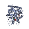 7yxdC 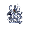 7yxnC 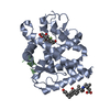 7yxoC  7yxpC 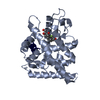 7yxrC 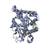 5ufsS S: Starting model for refinement C: citing same article ( |
|---|---|
| Similar structure data |
-Links
 PDBj
PDBj


- Assembly
| Deposited unit | 
| ||||||||
|---|---|---|---|---|---|---|---|---|---|
| 1 |
| ||||||||
| Unit cell |
| ||||||||
| Components on special symmetry positions |
|
-Components
-Protein / Protein/peptide , 2 types, 2 molecules AR
| #1: Protein | Mass: 28649.363 Da / Num. of mol.: 1 Source method: isolated from a genetically manipulated source Source: (gene. exp.) unidentified (others) / Production host:  |
|---|---|
| #2: Protein/peptide | Mass: 1204.440 Da / Num. of mol.: 1 Source method: isolated from a genetically manipulated source Source: (gene. exp.) Homo sapiens (human) / Gene: NR0B2, SHP / Production host: synthetic construct (others) / References: UniProt: Q15466 |
-Non-polymers , 4 types, 28 molecules 


| #3: Chemical | ChemComp-DEX /  Mass: 392.461 Da / Num. of mol.: 1 / Source method: obtained synthetically / Formula: C22H29FO5 / Feature type: SUBJECT OF INVESTIGATION Mass: 392.461 Da / Num. of mol.: 1 / Source method: obtained synthetically / Formula: C22H29FO5 / Feature type: SUBJECT OF INVESTIGATION | ||
|---|---|---|---|
| #4: Chemical | ChemComp-PG4 /  Mass: 194.226 Da / Num. of mol.: 1 / Source method: obtained synthetically / Formula: C8H18O5 / Comment: precipitant*YM Mass: 194.226 Da / Num. of mol.: 1 / Source method: obtained synthetically / Formula: C8H18O5 / Comment: precipitant*YM | ||
| #5: Chemical |  Mass: 60.009 Da / Num. of mol.: 2 / Source method: obtained synthetically / Formula: CO3 Mass: 60.009 Da / Num. of mol.: 2 / Source method: obtained synthetically / Formula: CO3#6: Water | ChemComp-HOH / | Mass: 18.015 Da / Num. of mol.: 24 / Source method: isolated from a natural source / Formula: H2O |
-Details
| Has ligand of interest | Y |
|---|
-Experimental details
-Experiment
| Experiment | Method: X-RAY DIFFRACTION / Number of used crystals: 1 |
|---|
- Sample preparation
| Crystal | Density Matthews: 2.39 Å3/Da / Density % sol: 48.47 % |
|---|---|
| Crystal grow | Temperature: 293.15 K / Method: vapor diffusion, sitting drop / pH: 6.5 Details: 85 mM sodium cacodylate trihydrate, pH 6.5, 0.17 M sodium acetate trihydrate, 25.5% (w/v) PEG8000, 15% (v/v) glycerol |
-Data collection
| Diffraction | Mean temperature: 100 K / Serial crystal experiment: N |
|---|---|
| Diffraction source | Source: SYNCHROTRON / Site: ALBA / Beamline: XALOC / Wavelength: 0.9788 Å |
| Detector | Type: DECTRIS PILATUS 6M / Detector: PIXEL / Date: Sep 30, 2018 |
| Radiation | Monochromator: M / Protocol: SINGLE WAVELENGTH / Monochromatic (M) / Laue (L): M / Scattering type: x-ray |
| Radiation wavelength | Wavelength: 0.9788 Å / Relative weight: 1 |
| Reflection | Resolution: 2.25→43.42 Å / Num. obs: 12530 / % possible obs: 93.8 % / Redundancy: 2.7 % / Biso Wilson estimate: 44.7 Å2 / CC1/2: 0.997 / Rpim(I) all: 0.039 / Rrim(I) all: 0.069 / Net I/σ(I): 11.1 |
| Reflection shell | Resolution: 2.25→2.32 Å / Redundancy: 2.2 % / Mean I/σ(I) obs: 1.8 / Num. unique obs: 907 / CC1/2: 0.654 / Rpim(I) all: 0.439 / Rrim(I) all: 0.697 / % possible all: 75.5 |
- Processing
| Software |
| |||||||||||||||||||||||||||||||||||||||||||||
|---|---|---|---|---|---|---|---|---|---|---|---|---|---|---|---|---|---|---|---|---|---|---|---|---|---|---|---|---|---|---|---|---|---|---|---|---|---|---|---|---|---|---|---|---|---|---|
| Refinement | Method to determine structure: MOLECULAR REPLACEMENT Starting model: 5UFS Resolution: 2.25→43.42 Å / Cor.coef. Fo:Fc: 0.961 / Cor.coef. Fo:Fc free: 0.942 / SU B: 5.412 / SU ML: 0.14 / Cross valid method: THROUGHOUT / σ(F): 0 / ESU R: 0.076 / ESU R Free: 0.051 / Stereochemistry target values: MAXIMUM LIKELIHOOD / Details: U VALUES : REFINED INDIVIDUALLY
| |||||||||||||||||||||||||||||||||||||||||||||
| Solvent computation | Ion probe radii: 0.8 Å / Shrinkage radii: 0.8 Å / VDW probe radii: 1.2 Å / Solvent model: MASK | |||||||||||||||||||||||||||||||||||||||||||||
| Displacement parameters | Biso max: 144.98 Å2 / Biso mean: 54.908 Å2 / Biso min: 25.09 Å2
| |||||||||||||||||||||||||||||||||||||||||||||
| Refinement step | Cycle: final / Resolution: 2.25→43.42 Å
| |||||||||||||||||||||||||||||||||||||||||||||
| Refine LS restraints |
| |||||||||||||||||||||||||||||||||||||||||||||
| LS refinement shell | Resolution: 2.25→2.309 Å / Rfactor Rfree error: 0 / Total num. of bins used: 20
|
