 Movie
Movie Controller


+ Open data

- Basic information
| Entry | Database: PDB / ID: 7yul | |||||||||
|---|---|---|---|---|---|---|---|---|---|---|
| Title | Crystal structure of human BEND6 BEN domain in complex with DNA | |||||||||
 Components Components |
| |||||||||
 Keywords Keywords | DNA BINDING PROTEIN / complex structure | |||||||||
| Function / homology |  Function and homology information Function and homology informationnegative regulation of Notch signaling pathway / positive regulation of neuron differentiation / transcription corepressor activity / nervous system development / chromatin binding / DNA-templated transcription / DNA binding / nucleus Similarity search - Function | |||||||||
| Biological species |  Homo sapiens (human) Homo sapiens (human)synthetic construct (others) | |||||||||
| Method |  X-RAY DIFFRACTION / MOLECULAR REPLACEMENT / Resolution: 1.82 Å X-RAY DIFFRACTION / MOLECULAR REPLACEMENT / Resolution: 1.82 Å | |||||||||
 Authors Authors | Liu, K. / Xiao, Y.Q. / Zhang, J. / Min, J.R. | |||||||||
| Funding support |  China, 1items China, 1items
| |||||||||
 Citation Citation | Journal: J.Biol.Chem. / Year: 2023 Title: Structural insights into DNA recognition by the BEN domain of the transcription factor BANP. Authors: Liu, K. / Zhang, J. / Xiao, Y. / Yang, A. / Song, X. / Li, Y. / Chen, Y. / Hughes, T.R. / Min, J. | |||||||||
| History |
|
- Structure visualization
| Structure viewer | Molecule: MolmilJmol/JSmol |
|---|
- Downloads & links
-Download
| PDBx/mmCIF format | 7yul.cif.gz | 47.7 KB | Display | PDBx/mmCIF format |
|---|---|---|---|---|
| PDB format | pdb7yul.ent.gz | 27.8 KB | Display | PDB format |
| PDBx/mmJSON format | 7yul.json.gz | Tree view | PDBx/mmJSON format | |
| Others |  Other downloads Other downloads |
-Validation report
| Arichive directory | https://data.pdbj.org/pub/pdb/validation_reports/yu/7yulftp://data.pdbj.org/pub/pdb/validation_reports/yu/7yul | HTTPS FTP |
|---|
-Related structure data
| Related structure data |  7yugSC  7yukC  7yunC  8htxC S: Starting model for refinement C: citing same article ( |
|---|---|
| Similar structure data |
-Links
 PDBj
PDBj


- Assembly
| Deposited unit | 
| |||||||||
|---|---|---|---|---|---|---|---|---|---|---|
| 1 | 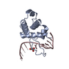
| |||||||||
| Unit cell |
| |||||||||
| Components on special symmetry positions |
|
-Components
| #1: Protein | Mass: 11806.545 Da / Num. of mol.: 1 Source method: isolated from a genetically manipulated source Source: (gene. exp.) Homo sapiens (human) / Gene: BEND6, C6orf65 / Production host:  |
|---|---|
| #2: DNA chain | Mass: 3663.392 Da / Num. of mol.: 1 Source method: isolated from a genetically manipulated source Source: (gene. exp.) synthetic construct (others) / Production host: synthetic construct (others) |
| #3: Chemical | ChemComp-GOA / 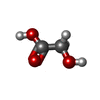  Mass: 76.051 Da / Num. of mol.: 1 / Source method: obtained synthetically / Formula: C2H4O3 Mass: 76.051 Da / Num. of mol.: 1 / Source method: obtained synthetically / Formula: C2H4O3 |
| #4: Water | ChemComp-HOH /  Mass: 18.015 Da / Num. of mol.: 140 / Source method: isolated from a natural source / Formula: H2O Mass: 18.015 Da / Num. of mol.: 140 / Source method: isolated from a natural source / Formula: H2O |
| Has ligand of interest | N |
-Experimental details
-Experiment
| Experiment | Method: X-RAY DIFFRACTION / Number of used crystals: 1 |
|---|
- Sample preparation
| Crystal | Density Matthews: 1.94 Å3/Da / Density % sol: 36.47 % |
|---|---|
| Crystal grow | Temperature: 291 K / Method: vapor diffusion, sitting drop Details: 0.12 M Ethylene glycol, 0.1 M Sodium HEPES, 0.1 M MOPS (acid), 12.5% MPD (v/v), 12.5% PEG 1000 (v/v), 12.5% PEG3350 (v/v) |
-Data collection
| Diffraction | Mean temperature: 100 K / Serial crystal experiment: N |
|---|---|
| Diffraction source | Source: LIQUID ANODE / Type: BRUKER METALJET / Wavelength: 1.34138 Å |
| Detector | Type: Bruker PHOTON III / Detector: PIXEL / Date: May 20, 2022 |
| Radiation | Protocol: SINGLE WAVELENGTH / Monochromatic (M) / Laue (L): M / Scattering type: x-ray |
| Radiation wavelength | Wavelength: 1.34138 Å / Relative weight: 1 |
| Reflection | Resolution: 1.82→27.11 Å / Num. obs: 11195 / % possible obs: 99.4 % / Redundancy: 7.69 % / Rmerge(I) obs: 0.0451 / Net I/σ(I): 34.15 |
| Reflection shell | Resolution: 1.82→1.85 Å / Redundancy: 3.26 % / Rmerge(I) obs: 0.159 / Mean I/σ(I) obs: 9.77 / Num. unique obs: 483 / % possible all: 92.7 |
- Processing
| Software |
| ||||||||||||||||||||||||||||||||||||||||||||||||||||||||||||
|---|---|---|---|---|---|---|---|---|---|---|---|---|---|---|---|---|---|---|---|---|---|---|---|---|---|---|---|---|---|---|---|---|---|---|---|---|---|---|---|---|---|---|---|---|---|---|---|---|---|---|---|---|---|---|---|---|---|---|---|---|---|
| Refinement | Method to determine structure: MOLECULAR REPLACEMENT Starting model: 7YUG Resolution: 1.82→27.11 Å / Cor.coef. Fo:Fc: 0.961 / Cor.coef. Fo:Fc free: 0.947 / SU B: 2.207 / SU ML: 0.069 / Cross valid method: THROUGHOUT / σ(F): 0 / ESU R: 0.127 / ESU R Free: 0.118 / Stereochemistry target values: MAXIMUM LIKELIHOOD Details: HYDROGENS HAVE BEEN ADDED IN THE RIDING POSITIONS U VALUES : REFINED INDIVIDUALLY
| ||||||||||||||||||||||||||||||||||||||||||||||||||||||||||||
| Solvent computation | Ion probe radii: 0.8 Å / Shrinkage radii: 0.8 Å / VDW probe radii: 1.2 Å / Solvent model: MASK | ||||||||||||||||||||||||||||||||||||||||||||||||||||||||||||
| Displacement parameters | Biso max: 85.64 Å2 / Biso mean: 16.862 Å2 / Biso min: 5.94 Å2
| ||||||||||||||||||||||||||||||||||||||||||||||||||||||||||||
| Refinement step | Cycle: final / Resolution: 1.82→27.11 Å
| ||||||||||||||||||||||||||||||||||||||||||||||||||||||||||||
| Refine LS restraints |
| ||||||||||||||||||||||||||||||||||||||||||||||||||||||||||||
| LS refinement shell | Resolution: 1.82→1.867 Å / Rfactor Rfree error: 0 / Total num. of bins used: 20
|
