 Movie
Movie Controller


+ Open data

- Basic information
| Entry | Database: PDB / ID: 7yuk | ||||||
|---|---|---|---|---|---|---|---|
| Title | Complex structure of BANP BEN domain bound to DNA | ||||||
 Components Components |
| ||||||
 Keywords Keywords | DNA BINDING PROTEIN / BANP-DNA complex structure | ||||||
| Function / homology |  Function and homology information Function and homology informationRegulation of TP53 Activity through Association with Co-factors / Degradation of CDH1 / chromatin organization / nuclear speck / nuclear body / DNA binding / RNA binding / nucleoplasm / identical protein binding / cytoplasm Similarity search - Function | ||||||
| Biological species |  Homo sapiens (human) Homo sapiens (human)synthetic construct (others) | ||||||
| Method |  X-RAY DIFFRACTION / SYNCHROTRON / MOLECULAR REPLACEMENT / Resolution: 2.11 Å X-RAY DIFFRACTION / SYNCHROTRON / MOLECULAR REPLACEMENT / Resolution: 2.11 Å | ||||||
 Authors Authors | Zhang, J. / Xiao, Y.Q. / Chen, Y.X. / Liu, K. / Min, J.R. | ||||||
| Funding support |  China, 1items China, 1items
| ||||||
 Citation Citation | Journal: J.Biol.Chem. / Year: 2023 Title: Structural insights into DNA recognition by the BEN domain of the transcription factor BANP. Authors: Liu, K. / Zhang, J. / Xiao, Y. / Yang, A. / Song, X. / Li, Y. / Chen, Y. / Hughes, T.R. / Min, J. | ||||||
| History |
|
- Structure visualization
| Structure viewer | Molecule: MolmilJmol/JSmol |
|---|
- Downloads & links
-Download
| PDBx/mmCIF format | 7yuk.cif.gz | 57 KB | Display | PDBx/mmCIF format |
|---|---|---|---|---|
| PDB format | pdb7yuk.ent.gz | 35.1 KB | Display | PDB format |
| PDBx/mmJSON format | 7yuk.json.gz | Tree view | PDBx/mmJSON format | |
| Others |  Other downloads Other downloads |
-Validation report
| Arichive directory | https://data.pdbj.org/pub/pdb/validation_reports/yu/7yukftp://data.pdbj.org/pub/pdb/validation_reports/yu/7yuk | HTTPS FTP |
|---|
-Related structure data
| Related structure data |  7yugSC 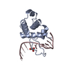 7yulC  7yunC  8htxC S: Starting model for refinement C: citing same article ( |
|---|---|
| Similar structure data |
-Links
 PDBj
PDBj


- Assembly
| Deposited unit | 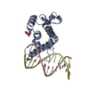
| ||||||||
|---|---|---|---|---|---|---|---|---|---|
| 1 |
| ||||||||
| Unit cell |
|
-Components
| #1: Protein | Mass: 16245.380 Da / Num. of mol.: 1 Source method: isolated from a genetically manipulated source Source: (gene. exp.) Homo sapiens (human) / Gene: BANP, BEND1, SMAR1 / Production host:  | ||||||
|---|---|---|---|---|---|---|---|
| #2: DNA chain | Mass: 3663.392 Da / Num. of mol.: 2 Source method: isolated from a genetically manipulated source Source: (gene. exp.) synthetic construct (others) / Production host: synthetic construct (others) #3: Chemical | ChemComp-GOL / |   Mass: 92.094 Da / Num. of mol.: 1 / Source method: isolated from a natural source / Formula: C3H8O3 Mass: 92.094 Da / Num. of mol.: 1 / Source method: isolated from a natural source / Formula: C3H8O3#4: Water | ChemComp-HOH / |  Mass: 18.015 Da / Num. of mol.: 55 / Source method: isolated from a natural source / Formula: H2O Mass: 18.015 Da / Num. of mol.: 55 / Source method: isolated from a natural source / Formula: H2OHas ligand of interest | N | |
-Experimental details
-Experiment
| Experiment | Method: X-RAY DIFFRACTION / Number of used crystals: 1 |
|---|
- Sample preparation
| Crystal | Density Matthews: 2.04 Å3/Da / Density % sol: 39.82 % |
|---|---|
| Crystal grow | Temperature: 291 K / Method: vapor diffusion, sitting drop Details: 20% w/v PEG3350; 0.02 M Sodium/potassium phosphate; 0.1 M Bris-Tris propane pH 6.5 |
-Data collection
| Diffraction | Mean temperature: 100 K / Serial crystal experiment: N |
|---|---|
| Diffraction source | Source: SYNCHROTRON / Site: SSRF / Beamline: BL18U1 / Wavelength: 0.9791 Å |
| Detector | Type: DECTRIS PILATUS3 6M / Detector: PIXEL / Date: Jan 13, 2022 |
| Radiation | Protocol: SINGLE WAVELENGTH / Monochromatic (M) / Laue (L): M / Scattering type: x-ray |
| Radiation wavelength | Wavelength: 0.9791 Å / Relative weight: 1 |
| Reflection | Resolution: 2.11→50 Å / Num. obs: 11802 / % possible obs: 99.9 % / Redundancy: 12.4 % / CC1/2: 0.998 / Rmerge(I) obs: 0.086 / Net I/σ(I): 17 |
| Reflection shell | Resolution: 2.11→2.17 Å / Redundancy: 12.2 % / Rmerge(I) obs: 0.471 / Mean I/σ(I) obs: 4.5 / Num. unique obs: 927 / CC1/2: 0.978 / % possible all: 99.4 |
- Processing
| Software |
| ||||||||||||||||||||||||||||||||||||||||||||||||||||||||||||
|---|---|---|---|---|---|---|---|---|---|---|---|---|---|---|---|---|---|---|---|---|---|---|---|---|---|---|---|---|---|---|---|---|---|---|---|---|---|---|---|---|---|---|---|---|---|---|---|---|---|---|---|---|---|---|---|---|---|---|---|---|---|
| Refinement | Method to determine structure: MOLECULAR REPLACEMENT Starting model: 7YUG Resolution: 2.11→40.17 Å / Cor.coef. Fo:Fc: 0.947 / Cor.coef. Fo:Fc free: 0.93 / SU B: 7.797 / SU ML: 0.189 / Cross valid method: THROUGHOUT / σ(F): 0 / ESU R: 0.259 / ESU R Free: 0.206 / Stereochemistry target values: MAXIMUM LIKELIHOOD Details: HYDROGENS HAVE BEEN ADDED IN THE RIDING POSITIONS U VALUES : REFINED INDIVIDUALLY
| ||||||||||||||||||||||||||||||||||||||||||||||||||||||||||||
| Solvent computation | Ion probe radii: 0.8 Å / Shrinkage radii: 0.8 Å / VDW probe radii: 1.2 Å / Solvent model: MASK | ||||||||||||||||||||||||||||||||||||||||||||||||||||||||||||
| Displacement parameters | Biso max: 81.92 Å2 / Biso mean: 40.692 Å2 / Biso min: 21.67 Å2
| ||||||||||||||||||||||||||||||||||||||||||||||||||||||||||||
| Refinement step | Cycle: final / Resolution: 2.11→40.17 Å
| ||||||||||||||||||||||||||||||||||||||||||||||||||||||||||||
| Refine LS restraints |
| ||||||||||||||||||||||||||||||||||||||||||||||||||||||||||||
| LS refinement shell | Resolution: 2.11→2.162 Å / Rfactor Rfree error: 0
|
