+ データを開く
- 基本情報
基本情報
| 登録情報 | データベース: PDB / ID: 7ymz | ||||||
|---|---|---|---|---|---|---|---|
| タイトル | Cryo-EM structure of MERS-CoV spike protein, intermediate conformation | ||||||
 要素 要素 | Spike glycoprotein | ||||||
 キーワード キーワード | VIRAL PROTEIN / MERS-CoV / Spike / Glycoprotein | ||||||
| 機能・相同性 |  機能・相同性情報 機能・相同性情報host cell endoplasmic reticulum-Golgi intermediate compartment membrane / membrane fusion / receptor-mediated endocytosis of virus by host cell / positive regulation of viral entry into host cell / receptor-mediated virion attachment to host cell / symbiont entry into host cell / fusion of virus membrane with host plasma membrane / fusion of virus membrane with host endosome membrane / viral envelope / host cell plasma membrane ...host cell endoplasmic reticulum-Golgi intermediate compartment membrane / membrane fusion / receptor-mediated endocytosis of virus by host cell / positive regulation of viral entry into host cell / receptor-mediated virion attachment to host cell / symbiont entry into host cell / fusion of virus membrane with host plasma membrane / fusion of virus membrane with host endosome membrane / viral envelope / host cell plasma membrane / virion membrane / membrane 類似検索 - 分子機能 | ||||||
| 生物種 |  Human betacoronavirus 2c EMC/2012 (ウイルス) Human betacoronavirus 2c EMC/2012 (ウイルス) | ||||||
| 手法 | 電子顕微鏡法 / 単粒子再構成法 / クライオ電子顕微鏡法 / 解像度: 4.39 Å | ||||||
 データ登録者 データ登録者 | Hsu, S.T.D. / Chang, N.E. / Weng, Z.W. / Yang, T.J. / Draczkowski, P. | ||||||
| 資金援助 |  台湾, 1件 台湾, 1件
| ||||||
 引用 引用 |  ジャーナル: Cell / 年: 2024 ジャーナル: Cell / 年: 2024タイトル: Rapid simulation of glycoprotein structures by grafting and steric exclusion of glycan conformer libraries. 著者: Yu-Xi Tsai / Ning-En Chang / Klaus Reuter / Hao-Ting Chang / Tzu-Jing Yang / Sören von Bülow / Vidhi Sehrawat / Noémie Zerrouki / Matthieu Tuffery / Michael Gecht / Isabell Louise Grothaus ...著者: Yu-Xi Tsai / Ning-En Chang / Klaus Reuter / Hao-Ting Chang / Tzu-Jing Yang / Sören von Bülow / Vidhi Sehrawat / Noémie Zerrouki / Matthieu Tuffery / Michael Gecht / Isabell Louise Grothaus / Lucio Colombi Ciacchi / Yong-Sheng Wang / Min-Feng Hsu / Kay-Hooi Khoo / Gerhard Hummer / Shang-Te Danny Hsu / Cyril Hanus / Mateusz Sikora /      要旨: Most membrane proteins are modified by covalent addition of complex sugars through N- and O-glycosylation. Unlike proteins, glycans do not typically adopt specific secondary structures and remain ...Most membrane proteins are modified by covalent addition of complex sugars through N- and O-glycosylation. Unlike proteins, glycans do not typically adopt specific secondary structures and remain very mobile, shielding potentially large fractions of protein surface. High glycan conformational freedom hinders complete structural elucidation of glycoproteins. Computer simulations may be used to model glycosylated proteins but require hundreds of thousands of computing hours on supercomputers, thus limiting routine use. Here, we describe GlycoSHIELD, a reductionist method that can be implemented on personal computers to graft realistic ensembles of glycan conformers onto static protein structures in minutes. Using molecular dynamics simulation, small-angle X-ray scattering, cryoelectron microscopy, and mass spectrometry, we show that this open-access toolkit provides enhanced models of glycoprotein structures. Focusing on N-cadherin, human coronavirus spike proteins, and gamma-aminobutyric acid receptors, we show that GlycoSHIELD can shed light on the impact of glycans on the conformation and activity of complex glycoproteins. | ||||||
| 履歴 |
|
- 構造の表示
構造の表示
| 構造ビューア | 分子:  Molmil Molmil Jmol/JSmol Jmol/JSmol |
|---|
- ダウンロードとリンク
- ダウンロード
| PDBx/mmCIF形式 |  7ymz.cif.gz 7ymz.cif.gz | 632.1 KB | 表示 |  PDBx/mmCIF形式 PDBx/mmCIF形式 |
|---|---|---|---|---|
| PDB形式 |  pdb7ymz.ent.gz pdb7ymz.ent.gz | 513.6 KB | 表示 |  PDB形式 PDB形式 |
| PDBx/mmJSON形式 |  7ymz.json.gz 7ymz.json.gz | ツリー表示 |  PDBx/mmJSON形式 PDBx/mmJSON形式 | |
| その他 |  その他のダウンロード その他のダウンロード |
-検証レポート
| 文書・要旨 |  7ymz_validation.pdf.gz 7ymz_validation.pdf.gz | 2.4 MB | 表示 |  wwPDB検証レポート wwPDB検証レポート |
|---|---|---|---|---|
| 文書・詳細版 |  7ymz_full_validation.pdf.gz 7ymz_full_validation.pdf.gz | 2.5 MB | 表示 | |
| XML形式データ |  7ymz_validation.xml.gz 7ymz_validation.xml.gz | 99.7 KB | 表示 | |
| CIF形式データ |  7ymz_validation.cif.gz 7ymz_validation.cif.gz | 155.5 KB | 表示 | |
| アーカイブディレクトリ |  https://data.pdbj.org/pub/pdb/validation_reports/ym/7ymz https://data.pdbj.org/pub/pdb/validation_reports/ym/7ymz ftp://data.pdbj.org/pub/pdb/validation_reports/ym/7ymz ftp://data.pdbj.org/pub/pdb/validation_reports/ym/7ymz | HTTPS FTP |
-関連構造データ
| 関連構造データ |  33948MC 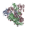 7ymtC 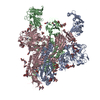 7ymvC 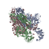 7ymwC 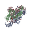 7ymxC 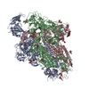 7ymyC  7yn0C M: このデータのモデリングに利用したマップデータ C: 同じ文献を引用 ( |
|---|---|
| 類似構造データ | 類似検索 - 機能・相同性  F&H 検索 F&H 検索 |
- リンク
リンク
- 集合体
集合体
| 登録構造単位 | 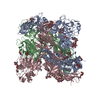
|
|---|---|
| 1 |
|
- 要素
要素
| #1: タンパク質 | 分子量: 150654.438 Da / 分子数: 3 / 変異: R748A, R751G, V1060P, L1061P / 由来タイプ: 組換発現 由来: (組換発現)  Human betacoronavirus 2c EMC/2012 (ウイルス) Human betacoronavirus 2c EMC/2012 (ウイルス)発現宿主:  Homo sapiens (ヒト) / 参照: UniProt: K0BRG7 Homo sapiens (ヒト) / 参照: UniProt: K0BRG7#2: 多糖 | 2-acetamido-2-deoxy-beta-D-glucopyranose-(1-4)-2-acetamido-2-deoxy-beta-D-glucopyranose #3: 多糖 | #4: 多糖 | alpha-L-fucopyranose-(1-6)-2-acetamido-2-deoxy-beta-D-glucopyranose #5: 糖 | ChemComp-NAG / 研究の焦点であるリガンドがあるか | Y | |
|---|
-実験情報
-実験
| 実験 | 手法: 電子顕微鏡法 |
|---|---|
| EM実験 | 試料の集合状態: PARTICLE / 3次元再構成法: 単粒子再構成法 |
- 試料調製
試料調製
| 構成要素 | 名称: recombinant MERS-CoV (betacoronavirus 2c EMC 2012) fm2P Spike タイプ: ORGANELLE OR CELLULAR COMPONENT / Entity ID: #1 / 由来: RECOMBINANT | ||||||||||||||||||||
|---|---|---|---|---|---|---|---|---|---|---|---|---|---|---|---|---|---|---|---|---|---|
| 分子量 | 実験値: NO | ||||||||||||||||||||
| 由来(天然) | 生物種:  Human betacoronavirus 2c EMC/2012 (ウイルス) Human betacoronavirus 2c EMC/2012 (ウイルス) | ||||||||||||||||||||
| 由来(組換発現) | 生物種:  Homo sapiens (ヒト) / 細胞: Expi293F Homo sapiens (ヒト) / 細胞: Expi293F | ||||||||||||||||||||
| 緩衝液 | pH: 7.6 | ||||||||||||||||||||
| 緩衝液成分 |
| ||||||||||||||||||||
| 試料 | 濃度: 0.5 mg/ml / 包埋: NO / シャドウイング: NO / 染色: NO / 凍結: YES | ||||||||||||||||||||
| 急速凍結 | 装置: FEI VITROBOT MARK IV / 凍結剤: ETHANE / 湿度: 100 % / 凍結前の試料温度: 277.15 K 詳細: blot for 2.5 seconds before plunging; blot force: -1; waiting time: 30s. |
- 電子顕微鏡撮影
| 実験機器 |  モデル: Talos Arctica / 画像提供: FEI Company |
|---|---|
| 顕微鏡 | モデル: FEI TALOS ARCTICA |
| 電子銃 | 電子線源:  FIELD EMISSION GUN / 加速電圧: 200 kV / 照射モード: FLOOD BEAM FIELD EMISSION GUN / 加速電圧: 200 kV / 照射モード: FLOOD BEAM |
| 電子レンズ | モード: BRIGHT FIELD / 倍率(公称値): 92000 X / 最大 デフォーカス(公称値): 1700 nm / 最小 デフォーカス(公称値): 1500 nm |
| 撮影 | 電子線照射量: 40.6 e/Å2 フィルム・検出器のモデル: FEI FALCON III (4k x 4k) 撮影したグリッド数: 1 / 実像数: 2886 |
- 解析
解析
| ソフトウェア | 名称: UCSF ChimeraX / バージョン: 1.2/v9 / 分類: モデル構築 / URL: https://www.rbvi.ucsf.edu/chimerax/ / Os: Windows / タイプ: package | ||||||||||||||||||||||||||||
|---|---|---|---|---|---|---|---|---|---|---|---|---|---|---|---|---|---|---|---|---|---|---|---|---|---|---|---|---|---|
| EMソフトウェア |
| ||||||||||||||||||||||||||||
| CTF補正 | タイプ: PHASE FLIPPING AND AMPLITUDE CORRECTION | ||||||||||||||||||||||||||||
| 粒子像の選択 | 選択した粒子像数: 1679870 | ||||||||||||||||||||||||||||
| 3次元再構成 | 解像度: 4.39 Å / 解像度の算出法: FSC 0.143 CUT-OFF / 粒子像の数: 58940 / 対称性のタイプ: POINT | ||||||||||||||||||||||||||||
| 原子モデル構築 | プロトコル: RIGID BODY FIT / 空間: REAL |
 ムービー
ムービー コントローラー


 PDBj
PDBj

