| 登録情報 | データベース: PDB / ID: 7ykg
|
|---|
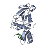
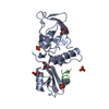
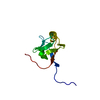
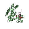
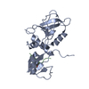
| タイトル | Crystal structure of MAGI2 PDZ0-GK/pSGEF complex |
|---|
 要素 要素 | - Membrane-associated guanylate kinase, WW and PDZ domain-containing protein 2
- SGEF
|
|---|
 キーワード キーワード | PEPTIDE BINDING PROTEIN / MAGI2 / PDZ0-GK / SGEF / phosphorylated peptide |
|---|
| 機能・相同性 |  機能・相同性情報 機能・相同性情報
type II activin receptor binding / neuroligin clustering involved in postsynaptic membrane assembly / podocyte development / slit diaphragm / positive regulation of synaptic vesicle clustering / beta-1 adrenergic receptor binding / activin receptor binding / nerve growth factor signaling pathway / SMAD protein signal transduction / clathrin-dependent endocytosis ...type II activin receptor binding / neuroligin clustering involved in postsynaptic membrane assembly / podocyte development / slit diaphragm / positive regulation of synaptic vesicle clustering / beta-1 adrenergic receptor binding / activin receptor binding / nerve growth factor signaling pathway / SMAD protein signal transduction / clathrin-dependent endocytosis / negative regulation of activin receptor signaling pathway / ciliary base / SMAD binding / receptor clustering / positive regulation of receptor internalization / photoreceptor outer segment / bicellular tight junction / phosphatase binding / photoreceptor inner segment / centriole / negative regulation of phosphatidylinositol 3-kinase/protein kinase B signal transduction / negative regulation of cell migration / positive regulation of neuron projection development / cellular response to nerve growth factor stimulus / late endosome / signaling receptor complex adaptor activity / postsynaptic density / negative regulation of cell population proliferation / dendrite / synapse / centrosome / perinuclear region of cytoplasm / signal transduction / protein-containing complex / nucleus / plasma membrane / cytoplasm類似検索 - 分子機能 Unstructured region on MAGI / Guanylate Kinase phosphate binding domain / Guanylate Kinase phosphate binding domain / Guanylate kinase, conserved site / Guanylate kinase-like signature. / Guanylate kinase-like domain profile. / Guanylate kinase-like domain / Guanylate kinase/L-type calcium channel beta subunit / Guanylate kinase / Guanylate kinase homologues. ...Unstructured region on MAGI / Guanylate Kinase phosphate binding domain / Guanylate Kinase phosphate binding domain / Guanylate kinase, conserved site / Guanylate kinase-like signature. / Guanylate kinase-like domain profile. / Guanylate kinase-like domain / Guanylate kinase/L-type calcium channel beta subunit / Guanylate kinase / Guanylate kinase homologues. / WW domain / WW/rsp5/WWP domain signature. / WW domain superfamily / WW/rsp5/WWP domain profile. / Domain with 2 conserved Trp (W) residues / WW domain / PDZ domain / PDZ domain profile. / Domain present in PSD-95, Dlg, and ZO-1/2. / PDZ domain / PDZ superfamily / P-loop containing nucleoside triphosphate hydrolase / 2-Layer Sandwich / Alpha Beta類似検索 - ドメイン・相同性 Membrane-associated guanylate kinase, WW and PDZ domain-containing protein 2類似検索 - 構成要素 |
|---|
| 生物種 |   Mus musculus (ハツカネズミ) Mus musculus (ハツカネズミ) |
|---|
| 手法 |  X線回折 / シンクロトロン / 分子置換 / 解像度: 2.16 Å X線回折 / シンクロトロン / 分子置換 / 解像度: 2.16 Å |
|---|
 データ登録者 データ登録者 | Zhang, M. / Lin, L. / Zhu, J. |
|---|
| 資金援助 | 1件 |
|---|
 引用 引用 | ジャーナル: Sci Adv / 年: 2023
タイトル: Phosphorylation-dependent recognition of diverse protein targets by the cryptic GK domain of MAGI MAGUKs.
著者: Zhang, M. / Cao, A. / Lin, L. / Chen, Y. / Shang, Y. / Wang, C. / Zhang, M. / Zhu, J. |
|---|
| 履歴 | | 登録 | 2022年7月22日 | 登録サイト: PDBJ / 処理サイト: PDBJ |
|---|
| 改定 1.0 | 2023年8月2日 | Provider: repository / タイプ: Initial release |
|---|
| 改定 1.1 | 2024年2月28日 | Group: Data collection / Database references
カテゴリ: chem_comp_atom / chem_comp_bond ...chem_comp_atom / chem_comp_bond / citation / citation_author
Item: _citation.country / _citation.journal_abbrev ..._citation.country / _citation.journal_abbrev / _citation.journal_id_CSD / _citation.journal_id_ISSN / _citation.journal_volume / _citation.page_first / _citation.page_last / _citation.pdbx_database_id_DOI / _citation.pdbx_database_id_PubMed / _citation.title / _citation.year |
|---|
| 改定 1.2 | 2024年11月20日 | Group: Structure summary
カテゴリ: pdbx_entry_details / pdbx_modification_feature
Item: _pdbx_entry_details.has_protein_modification |
|---|
|
|---|
 ムービー
ムービー コントローラー


 データを開く
 基本情報
基本情報 構造の表示
構造の表示 ダウンロードとリンク
 その他のダウンロード
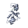
 PDBj
PDBj
 集合体
集合体


 分子量: 18.015 Da / 分子数: 224 / 由来タイプ: 天然 / 式: H2O
 試料調製
試料調製 / ビームライン: BL19U1 / 波長: 0.97737 Å
 解析
解析