 Movie
Movie Controller

[English] 日本語
 Yorodumi
- PDB-7yiv: The Crystal Structure of Human Tissue Nonspecific Alkaline Phosph... -
+ Open data

- Basic information
| Entry | Database: PDB / ID: 7yiv | ||||||
|---|---|---|---|---|---|---|---|
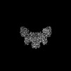
| Title | The Crystal Structure of Human Tissue Nonspecific Alkaline Phosphatase (ALPL) at Basic pH | ||||||
 Components Components | Alkaline phosphatase, tissue-nonspecific isozyme | ||||||
 Keywords Keywords | HYDROLASE / alkaline phosphatase / bone mineralization / catalytic network / hypophosphatasia / structural biology | ||||||
| Function / homology |  Function and homology information Function and homology informationphosphoamidase / pyridoxal 5'-phosphate metabolic process / phosphoamidase activity / phosphoethanolamine phosphatase activity / response to vitamin B6 / futile creatine cycle / pyridoxal phosphatase activity / developmental process involved in reproduction / ADP phosphatase activity / pyrophosphatase activity ...phosphoamidase / pyridoxal 5'-phosphate metabolic process / phosphoamidase activity / phosphoethanolamine phosphatase activity / response to vitamin B6 / futile creatine cycle / pyridoxal phosphatase activity / developmental process involved in reproduction / ADP phosphatase activity / pyrophosphatase activity / inhibition of non-skeletal tissue mineralization / response to macrophage colony-stimulating factor / cementum mineralization / Post-translational modification: synthesis of GPI-anchored proteins / alkaline phosphatase / alkaline phosphatase activity / response to sodium phosphate / inorganic diphosphate phosphatase activity / phosphate ion homeostasis / endochondral ossification / cellular homeostasis / response to vitamin D / bone mineralization / side of membrane / calcium ion homeostasis / response to glucocorticoid / skeletal system development / response to insulin / mitochondrial intermembrane space / mitochondrial membrane / osteoblast differentiation / positive regulation of cold-induced thermogenesis / extracellular matrix / response to lipopolysaccharide / response to antibiotic / calcium ion binding / ATP hydrolysis activity / extracellular exosome / extracellular region / membrane / plasma membrane Similarity search - Function | ||||||
| Biological species |  Homo sapiens (human) Homo sapiens (human) | ||||||
| Method |  X-RAY DIFFRACTION / SYNCHROTRON / MOLECULAR REPLACEMENT / Resolution: 3.18 Å X-RAY DIFFRACTION / SYNCHROTRON / MOLECULAR REPLACEMENT / Resolution: 3.18 Å | ||||||
 Authors Authors | Cao, Y. / Qin, A. / Yu, Y.T. / Yao, D.Q. / Zhang, Q. / Rao, B. / Xia, Y. / Lu, Y. | ||||||
| Funding support |  China, 1items China, 1items
| ||||||
 Citation Citation | Journal: Nat Commun / Year: 2023 Title: The structural pathology for hypophosphatasia caused by malfunctional tissue non-specific alkaline phosphatase. Authors: Yating Yu / Kewei Rong / Deqiang Yao / Qing Zhang / Xiankun Cao / Bing Rao / Ying Xia / Yi Lu / Yafeng Shen / Ying Yao / Hongtao Xu / Peixiang Ma / Yu Cao / An Qin / Abstract: Hypophosphatasia (HPP) is a metabolic bone disease that manifests as developmental abnormalities in bone and dental tissues. HPP patients exhibit hypo-mineralization and osteopenia due to the ...Hypophosphatasia (HPP) is a metabolic bone disease that manifests as developmental abnormalities in bone and dental tissues. HPP patients exhibit hypo-mineralization and osteopenia due to the deficiency or malfunction of tissue non-specific alkaline phosphatase (TNAP), which catalyzes the hydrolysis of phosphate-containing molecules outside the cells, promoting the deposition of hydroxyapatite in the extracellular matrix. Despite the identification of hundreds of pathogenic TNAP mutations, the detailed molecular pathology of HPP remains unclear. Here, to address this issue, we determine the crystal structures of human TNAP at near-atomic resolution and map the major pathogenic mutations onto the structure. Our study reveals an unexpected octameric architecture for TNAP, which is generated by the tetramerization of dimeric TNAPs, potentially stabilizing the TNAPs in the extracellular environments. Moreover, we use cryo-electron microscopy to demonstrate that the TNAP agonist antibody (JTALP001) forms a stable complex with TNAP by binding to the octameric interface. The administration of JTALP001 enhances osteoblast mineralization and promoted recombinant TNAP-rescued mineralization in TNAP knockout osteoblasts. Our findings elucidate the structural pathology of HPP and highlight the therapeutic potential of the TNAP agonist antibody for osteoblast-associated bone disorders. | ||||||
| History |
|
- Structure visualization
| Structure viewer | Molecule: MolmilJmol/JSmol |
|---|
- Downloads & links
-Download
| PDBx/mmCIF format | 7yiv.cif.gz | 749.1 KB | Display | PDBx/mmCIF format |
|---|---|---|---|---|
| PDB format | pdb7yiv.ent.gz | 619.6 KB | Display | PDB format |
| PDBx/mmJSON format | 7yiv.json.gz | Tree view | PDBx/mmJSON format | |
| Others |  Other downloads Other downloads |
-Validation report
| Arichive directory | https://data.pdbj.org/pub/pdb/validation_reports/yi/7yivftp://data.pdbj.org/pub/pdb/validation_reports/yi/7yiv | HTTPS FTP |
|---|
-Related structure data
| Related structure data | 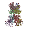 7yiwC 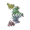 7yixC 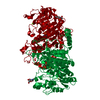 1ew2S S: Starting model for refinement C: citing same article ( |
|---|---|
| Similar structure data |
-Links
 PDBj
PDBj
- Assembly
| Deposited unit | 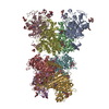
| ||||||||
|---|---|---|---|---|---|---|---|---|---|
| 1 |
| ||||||||
| Unit cell |
|
-Components
-Protein , 1 types, 8 molecules ADCBGHFE
| #1: Protein | Mass: 57020.188 Da / Num. of mol.: 8 Source method: isolated from a genetically manipulated source Source: (gene. exp.) Homo sapiens (human) / Gene: ALPL / Production host:  Trichoplusia ni (cabbage looper) Trichoplusia ni (cabbage looper)References: UniProt: P05186, alkaline phosphatase, phosphoamidase |
|---|
-Sugars , 2 types, 32 molecules 
| #2: Polysaccharide | 2-acetamido-2-deoxy-beta-D-glucopyranose-(1-4)-2-acetamido-2-deoxy-beta-D-glucopyranose Source method: isolated from a genetically manipulated source #3: Sugar | ChemComp-NAG /  Type: D-saccharide, beta linking / Mass: 221.208 Da / Num. of mol.: 16 / Source method: obtained synthetically / Formula: C8H15NO6 / Feature type: SUBJECT OF INVESTIGATION Type: D-saccharide, beta linking / Mass: 221.208 Da / Num. of mol.: 16 / Source method: obtained synthetically / Formula: C8H15NO6 / Feature type: SUBJECT OF INVESTIGATION |
|---|
-Non-polymers , 3 types, 32 molecules 


| #4: Chemical | ChemComp-MG /  Mass: 24.305 Da / Num. of mol.: 8 / Source method: obtained synthetically / Formula: Mg / Feature type: SUBJECT OF INVESTIGATION Mass: 24.305 Da / Num. of mol.: 8 / Source method: obtained synthetically / Formula: Mg / Feature type: SUBJECT OF INVESTIGATION#5: Chemical | ChemComp-ZN /  Mass: 65.409 Da / Num. of mol.: 16 / Source method: obtained synthetically / Formula: Zn / Feature type: SUBJECT OF INVESTIGATION Mass: 65.409 Da / Num. of mol.: 16 / Source method: obtained synthetically / Formula: Zn / Feature type: SUBJECT OF INVESTIGATION#6: Chemical | ChemComp-CA /  Mass: 40.078 Da / Num. of mol.: 8 / Source method: obtained synthetically / Formula: Ca / Feature type: SUBJECT OF INVESTIGATION Mass: 40.078 Da / Num. of mol.: 8 / Source method: obtained synthetically / Formula: Ca / Feature type: SUBJECT OF INVESTIGATION |
|---|
-Details
| Has ligand of interest | Y |
|---|---|
| Has protein modification | Y |
-Experimental details
-Experiment
| Experiment | Method: X-RAY DIFFRACTION / Number of used crystals: 1 |
|---|
- Sample preparation
| Crystal | Density Matthews: 2.75 Å3/Da / Density % sol: 55.21 % |
|---|---|
| Crystal grow | Temperature: 293 K / Method: vapor diffusion, sitting drop / pH: 8 Details: 17% (w/v) PEG4000, 30 mM Tris, pH 8.0, 100 mM MgCl2 |
-Data collection
| Diffraction | Mean temperature: 77 K / Serial crystal experiment: N |
|---|---|
| Diffraction source | Source: SYNCHROTRON / Site: SSRF / Beamline: BL18U1 / Wavelength: 1 Å |
| Detector | Type: DECTRIS PILATUS3 6M / Detector: PIXEL / Date: Jan 31, 2021 |
| Radiation | Protocol: SINGLE WAVELENGTH / Monochromatic (M) / Laue (L): M / Scattering type: x-ray |
| Radiation wavelength | Wavelength: 1 Å / Relative weight: 1 |
| Reflection | Resolution: 3.17→43.49 Å / Num. obs: 83898 / % possible obs: 98.62 % / Redundancy: 6.7 % / Biso Wilson estimate: 70.29 Å2 / CC1/2: 0.98 / Net I/σ(I): 2.28 |
| Reflection shell | Resolution: 3.2→3.26 Å / Redundancy: 6.5 % / Num. unique obs: 11103 / CC1/2: 0.663 / % possible all: 98.5 |
- Processing
| Software |
| |||||||||||||||||||||||||||||||||||||||||||||||||||||||||||||||||||||||||||||||||||||||||||||||||||||||||
|---|---|---|---|---|---|---|---|---|---|---|---|---|---|---|---|---|---|---|---|---|---|---|---|---|---|---|---|---|---|---|---|---|---|---|---|---|---|---|---|---|---|---|---|---|---|---|---|---|---|---|---|---|---|---|---|---|---|---|---|---|---|---|---|---|---|---|---|---|---|---|---|---|---|---|---|---|---|---|---|---|---|---|---|---|---|---|---|---|---|---|---|---|---|---|---|---|---|---|---|---|---|---|---|---|---|---|
| Refinement | Method to determine structure: MOLECULAR REPLACEMENT Starting model: 1EW2 Resolution: 3.18→25.05 Å / SU ML: 0.4 / Cross valid method: THROUGHOUT / σ(F): 1.34 / Phase error: 24.36 / Stereochemistry target values: ML
| |||||||||||||||||||||||||||||||||||||||||||||||||||||||||||||||||||||||||||||||||||||||||||||||||||||||||
| Solvent computation | Shrinkage radii: 0.9 Å / VDW probe radii: 1.11 Å / Solvent model: FLAT BULK SOLVENT MODEL | |||||||||||||||||||||||||||||||||||||||||||||||||||||||||||||||||||||||||||||||||||||||||||||||||||||||||
| Displacement parameters | Biso max: 137.86 Å2 / Biso mean: 68.6681 Å2 / Biso min: 28.54 Å2 | |||||||||||||||||||||||||||||||||||||||||||||||||||||||||||||||||||||||||||||||||||||||||||||||||||||||||
| Refinement step | Cycle: final / Resolution: 3.18→25.05 Å
| |||||||||||||||||||||||||||||||||||||||||||||||||||||||||||||||||||||||||||||||||||||||||||||||||||||||||
| LS refinement shell | Refine-ID: X-RAY DIFFRACTION / Rfactor Rfree error: 0 / Total num. of bins used: 14
|
