 Movie
Movie Controller


+ Open data

- Basic information
| Entry | Database: PDB / ID: 7ydt | ||||||
|---|---|---|---|---|---|---|---|
| Title | Crystal structure of mouse MPND | ||||||
 Components Components | MPN domain containing protein | ||||||
 Keywords Keywords | DNA BINDING PROTEIN / MPND | ||||||
| Function / homology |  Function and homology information Function and homology informationHydrolases; Acting on peptide bonds (peptidases) / metallopeptidase activity / proteolysis / metal ion binding Similarity search - Function | ||||||
| Biological species |  | ||||||
| Method |  X-RAY DIFFRACTION / SYNCHROTRON / MOLECULAR REPLACEMENT / Resolution: 2.055 Å X-RAY DIFFRACTION / SYNCHROTRON / MOLECULAR REPLACEMENT / Resolution: 2.055 Å | ||||||
 Authors Authors | Yang, M. / Chen, Z. | ||||||
| Funding support |  China, 1items China, 1items
| ||||||
 Citation Citation | Journal: Int J Mol Sci / Year: 2023 Title: Structures of MPND Reveal the Molecular Recognition of Nucleosomes. Authors: Yang, M. / Li, X. / Tian, Z. / Ma, L. / Ma, J. / Liu, Y. / Shang, G. / Liang, A. / Wu, W. / Chen, Z. | ||||||
| History |
|
- Structure visualization
| Structure viewer | Molecule: MolmilJmol/JSmol |
|---|
- Downloads & links
-Download
| PDBx/mmCIF format | 7ydt.cif.gz | 54.2 KB | Display | PDBx/mmCIF format |
|---|---|---|---|---|
| PDB format | pdb7ydt.ent.gz | 34.8 KB | Display | PDB format |
| PDBx/mmJSON format | 7ydt.json.gz | Tree view | PDBx/mmJSON format | |
| Others |  Other downloads Other downloads |
-Validation report
| Summary document | 7ydt_validation.pdf.gz | 426.4 KB | Display | wwPDB validaton report |
|---|---|---|---|---|
| Full document | 7ydt_full_validation.pdf.gz | 426.4 KB | Display | |
| Data in XML | 7ydt_validation.xml.gz | 8.8 KB | Display | |
| Data in CIF | 7ydt_validation.cif.gz | 11.6 KB | Display | |
| Arichive directory | https://data.pdbj.org/pub/pdb/validation_reports/yd/7ydtftp://data.pdbj.org/pub/pdb/validation_reports/yd/7ydt | HTTPS FTP |
-Related structure data
| Related structure data | 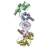 7ydwC 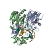 6pbdS S: Starting model for refinement C: citing same article ( |
|---|---|
| Similar structure data |
-Links
 PDBj
PDBj

- Assembly
| Deposited unit | 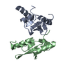
| ||||||||
|---|---|---|---|---|---|---|---|---|---|
| 1 | 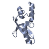
| ||||||||
| 2 | 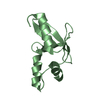
| ||||||||
| Unit cell |
|
-Components
| #1: Protein | Mass: 17005.990 Da / Num. of mol.: 2 Source method: isolated from a genetically manipulated source Source: (gene. exp.)  #2: Water | ChemComp-HOH / |  Mass: 18.015 Da / Num. of mol.: 62 / Source method: isolated from a natural source / Formula: H2O Mass: 18.015 Da / Num. of mol.: 62 / Source method: isolated from a natural source / Formula: H2O |
|---|
-Experimental details
-Experiment
| Experiment | Method: X-RAY DIFFRACTION / Number of used crystals: 1 |
|---|
- Sample preparation
| Crystal | Density Matthews: 1.96 Å3/Da / Density % sol: 37.24 % / Description: slice |
|---|---|
| Crystal grow | Temperature: 277 K / Method: vapor diffusion, sitting drop / Details: PEG4000, Bis Tris, ammonium acetate |
-Data collection
| Diffraction | Mean temperature: 100 K / Serial crystal experiment: N |
|---|---|
| Diffraction source | Source: SYNCHROTRON / Site: SSRF / Beamline: BL18U1 / Wavelength: 0.9793 Å |
| Detector | Type: DECTRIS PILATUS 2M / Detector: PIXEL / Date: Oct 26, 2020 |
| Radiation | Protocol: SINGLE WAVELENGTH / Monochromatic (M) / Laue (L): M / Scattering type: x-ray |
| Radiation wavelength | Wavelength: 0.9793 Å / Relative weight: 1 |
| Reflection | Resolution: 2.05→50 Å / Num. obs: 17365 / % possible obs: 97.6 % / Redundancy: 5.8 % / Rmerge(I) obs: 0.098 / Net I/σ(I): 13.3 |
| Reflection shell | Resolution: 2.05→2.1 Å / Num. unique obs: 732 / CC1/2: 0.883 |
- Processing
| Software |
| ||||||||||||||||||||||||||||||||||||||||||||||||||||||||||||||||||||||||||||||||||||||||||||||||||||||||||||||||||||||||||||||||||||||||||||||||||||||
|---|---|---|---|---|---|---|---|---|---|---|---|---|---|---|---|---|---|---|---|---|---|---|---|---|---|---|---|---|---|---|---|---|---|---|---|---|---|---|---|---|---|---|---|---|---|---|---|---|---|---|---|---|---|---|---|---|---|---|---|---|---|---|---|---|---|---|---|---|---|---|---|---|---|---|---|---|---|---|---|---|---|---|---|---|---|---|---|---|---|---|---|---|---|---|---|---|---|---|---|---|---|---|---|---|---|---|---|---|---|---|---|---|---|---|---|---|---|---|---|---|---|---|---|---|---|---|---|---|---|---|---|---|---|---|---|---|---|---|---|---|---|---|---|---|---|---|---|---|---|---|---|
| Refinement | Method to determine structure: MOLECULAR REPLACEMENT Starting model: 6PBD Resolution: 2.055→41.971 Å / Cor.coef. Fo:Fc: 0.937 / Cor.coef. Fo:Fc free: 0.929 / WRfactor Rfree: 0.249 / WRfactor Rwork: 0.23 / SU B: 4.897 / SU ML: 0.126 / Average fsc free: 0.8886 / Average fsc work: 0.8981 / Cross valid method: FREE R-VALUE / ESU R: 0.186 / ESU R Free: 0.16 Details: Hydrogens have been added in their riding positions
| ||||||||||||||||||||||||||||||||||||||||||||||||||||||||||||||||||||||||||||||||||||||||||||||||||||||||||||||||||||||||||||||||||||||||||||||||||||||
| Solvent computation | Ion probe radii: 0.8 Å / Shrinkage radii: 0.8 Å / VDW probe radii: 1.2 Å / Solvent model: MASK BULK SOLVENT | ||||||||||||||||||||||||||||||||||||||||||||||||||||||||||||||||||||||||||||||||||||||||||||||||||||||||||||||||||||||||||||||||||||||||||||||||||||||
| Displacement parameters | Biso mean: 34.645 Å2
| ||||||||||||||||||||||||||||||||||||||||||||||||||||||||||||||||||||||||||||||||||||||||||||||||||||||||||||||||||||||||||||||||||||||||||||||||||||||
| Refinement step | Cycle: LAST / Resolution: 2.055→41.971 Å
| ||||||||||||||||||||||||||||||||||||||||||||||||||||||||||||||||||||||||||||||||||||||||||||||||||||||||||||||||||||||||||||||||||||||||||||||||||||||
| Refine LS restraints |
| ||||||||||||||||||||||||||||||||||||||||||||||||||||||||||||||||||||||||||||||||||||||||||||||||||||||||||||||||||||||||||||||||||||||||||||||||||||||
| LS refinement shell |
|
