 Movie
Movie Controller

[English] 日本語
 Yorodumi
- PDB-7ycw: Crystal Form 1 of Truncated Antitoxin ParD (2-54,containg RHH dom... -
+ Open data

- Basic information
| Entry | Database: PDB / ID: 7ycw | ||||||
|---|---|---|---|---|---|---|---|
| Title | Crystal Form 1 of Truncated Antitoxin ParD (2-54,containg RHH domain) from Pseudoalteromonas rubra | ||||||
 Components Components | Antitoxin ParD | ||||||
 Keywords Keywords | ANTITOXIN / RHH / Transcription Factor / Toxin antitoxin system | ||||||
| Function / homology | Bacterial antitoxin of ParD toxin-antitoxin type II system and RHH / Antitoxin ParD / Antitoxin ParD superfamily / Ribbon-helix-helix / regulation of DNA-templated transcription / Antitoxin ParD Function and homology information Function and homology information | ||||||
| Biological species |  Pseudoalteromonas rubra (bacteria) Pseudoalteromonas rubra (bacteria) | ||||||
| Method |  X-RAY DIFFRACTION / SYNCHROTRON / MOLECULAR REPLACEMENT / Resolution: 2.2 Å X-RAY DIFFRACTION / SYNCHROTRON / MOLECULAR REPLACEMENT / Resolution: 2.2 Å | ||||||
 Authors Authors | Wang, C.C. / Niu, C.Y. / Niu, L.W. | ||||||
| Funding support |  China, 1items China, 1items
| ||||||
 Citation Citation | Journal: Front Microbiol / Year: 2022 Title: Structural insights into the PrpTA toxin-antitoxin system in Pseudoalteromonas rubra. Authors: Wang, C. / Niu, C. / Hidayatullah, K.M. / Xue, L. / Zhu, Z. / Niu, L. | ||||||
| History |
|
- Structure visualization
| Structure viewer | Molecule: MolmilJmol/JSmol |
|---|
- Downloads & links
-Download
| PDBx/mmCIF format | 7ycw.cif.gz | 55.8 KB | Display | PDBx/mmCIF format |
|---|---|---|---|---|
| PDB format | pdb7ycw.ent.gz | 38.7 KB | Display | PDB format |
| PDBx/mmJSON format | 7ycw.json.gz | Tree view | PDBx/mmJSON format | |
| Others |  Other downloads Other downloads |
-Validation report
| Arichive directory | https://data.pdbj.org/pub/pdb/validation_reports/yc/7ycwftp://data.pdbj.org/pub/pdb/validation_reports/yc/7ycw | HTTPS FTP |
|---|
-Related structure data
| Related structure data |  7ycsC  7ycuC  7ycvC 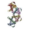 7b22S S: Starting model for refinement C: citing same article ( |
|---|---|
| Similar structure data |
-Links
 PDBj
PDBj- Assembly
| Deposited unit | 
| ||||||||||||
|---|---|---|---|---|---|---|---|---|---|---|---|---|---|
| 1 | 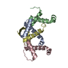
| ||||||||||||
| Unit cell |
|
-Components
| #1: Protein | Mass: 7066.906 Da / Num. of mol.: 4 Source method: isolated from a genetically manipulated source Source: (gene. exp.) Pseudoalteromonas rubra (bacteria) / Gene: AT705_24525 / Production host: #2: Water | ChemComp-HOH / |  Mass: 18.015 Da / Num. of mol.: 66 / Source method: isolated from a natural source / Formula: H2O Mass: 18.015 Da / Num. of mol.: 66 / Source method: isolated from a natural source / Formula: H2O |
|---|
-Experimental details
-Experiment
| Experiment | Method: X-RAY DIFFRACTION / Number of used crystals: 1 |
|---|
- Sample preparation
| Crystal | Density Matthews: 2.2 Å3/Da / Density % sol: 44.03 % |
|---|---|
| Crystal grow | Temperature: 289 K / Method: vapor diffusion, sitting drop / pH: 8 Details: 0.2 M Sodium acetate trihydrate,0.1 M Sodium citrate pH 5.5, 5 % w/v PEG 4000 |
-Data collection
| Diffraction | Mean temperature: 100 K / Serial crystal experiment: N |
|---|---|
| Diffraction source | Source: SYNCHROTRON / Site: SSRF / Beamline: BL19U1 / Wavelength: 0.97915 Å |
| Detector | Type: DECTRIS PILATUS 6M / Detector: PIXEL / Date: Dec 8, 2021 |
| Radiation | Protocol: SINGLE WAVELENGTH / Monochromatic (M) / Laue (L): M / Scattering type: x-ray |
| Radiation wavelength | Wavelength: 0.97915 Å / Relative weight: 1 |
| Reflection | Resolution: 2.2→30.78 Å / Num. obs: 13454 / % possible obs: 99.71 % / Observed criterion σ(I): 3 / Redundancy: 24.1 % / Biso Wilson estimate: 43.48 Å2 / CC1/2: 0.999 / CC star: 1 / Rmerge(I) obs: 0.06929 / Rpim(I) all: 0.01471 / Rrim(I) all: 0.07089 / Net I/σ(I): 30.41 |
| Reflection shell | Resolution: 2.2→2.279 Å / Redundancy: 25.8 % / Rmerge(I) obs: 0.601 / Mean I/σ(I) obs: 5.95 / Num. unique obs: 1294 / CC1/2: 0.989 / CC star: 0.997 / Rpim(I) all: 0.1194 / Rrim(I) all: 0.613 / % possible all: 98.92 |
- Processing
| Software |
| ||||||||||||||||||||||||||||||||||||||||||
|---|---|---|---|---|---|---|---|---|---|---|---|---|---|---|---|---|---|---|---|---|---|---|---|---|---|---|---|---|---|---|---|---|---|---|---|---|---|---|---|---|---|---|---|
| Refinement | Method to determine structure: MOLECULAR REPLACEMENT Starting model: 7B22 Resolution: 2.2→30.78 Å / SU ML: 0.2369 / Cross valid method: FREE R-VALUE / σ(F): 1.34 / Phase error: 28.1627 Stereochemistry target values: GeoStd + Monomer Library + CDL v1.2
| ||||||||||||||||||||||||||||||||||||||||||
| Solvent computation | Shrinkage radii: 0.9 Å / VDW probe radii: 1.1 Å / Solvent model: FLAT BULK SOLVENT MODEL | ||||||||||||||||||||||||||||||||||||||||||
| Displacement parameters | Biso mean: 52.42 Å2 | ||||||||||||||||||||||||||||||||||||||||||
| Refinement step | Cycle: LAST / Resolution: 2.2→30.78 Å
| ||||||||||||||||||||||||||||||||||||||||||
| Refine LS restraints |
| ||||||||||||||||||||||||||||||||||||||||||
| LS refinement shell |
|
