 Movie
Movie Controller


+ Open data

- Basic information
| Entry | Database: PDB / ID: 7b22 | ||||||
|---|---|---|---|---|---|---|---|
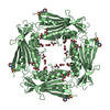
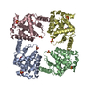
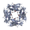
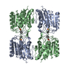
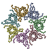
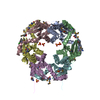
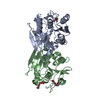
| Title | Vibrio cholerae ParD2 Antitoxin | ||||||
 Components Components | Antitoxin ParD | ||||||
 Keywords Keywords | DNA BINDING PROTEIN / Prokaryotic Toxin-Antitoxin System / intrinsically disordered proteins / RHH protein / transcriptional repressor / Antitoxin | ||||||
| Function / homology | Bacterial antitoxin of ParD toxin-antitoxin type II system and RHH / Antitoxin ParD / Antitoxin ParD superfamily / detoxification / toxin sequestering activity / Ribbon-helix-helix / regulation of DNA-templated transcription / Antitoxin ParD Function and homology information Function and homology information | ||||||
| Biological species |  Vibrio cholerae serotype O1 (bacteria) Vibrio cholerae serotype O1 (bacteria) | ||||||
| Method |  X-RAY DIFFRACTION / SYNCHROTRON / MOLECULAR REPLACEMENT / molecular replacement / Resolution: 3.08 Å X-RAY DIFFRACTION / SYNCHROTRON / MOLECULAR REPLACEMENT / molecular replacement / Resolution: 3.08 Å | ||||||
 Authors Authors | Garcia-Rodriguez, G. / Loris, R. | ||||||
 Citation Citation | Journal: Acta Crystallogr D Struct Biol / Year: 2021 Title: Entropic pressure controls the oligomerization of the Vibrio cholerae ParD2 antitoxin. Authors: Garcia-Rodriguez, G. / Girardin, Y. / Volkov, A.N. / Singh, R.K. / Muruganandam, G. / Van Dyck, J. / Sobott, F. / Versees, W. / Charlier, D. / Loris, R. | ||||||
| History |
|
- Structure visualization
| Structure viewer | Molecule: MolmilJmol/JSmol |
|---|
- Downloads & links
-Download
| PDBx/mmCIF format | 7b22.cif.gz | 82.9 KB | Display | PDBx/mmCIF format |
|---|---|---|---|---|
| PDB format | pdb7b22.ent.gz | 62.8 KB | Display | PDB format |
| PDBx/mmJSON format | 7b22.json.gz | Tree view | PDBx/mmJSON format | |
| Others |  Other downloads Other downloads |
-Validation report
| Arichive directory | https://data.pdbj.org/pub/pdb/validation_reports/b2/7b22ftp://data.pdbj.org/pub/pdb/validation_reports/b2/7b22 | HTTPS FTP |
|---|
-Related structure data
| Related structure data |  3kxeS S: Starting model for refinement |
|---|---|
| Similar structure data |

-Links
 PDBj
PDBj- Assembly
| Deposited unit | 
| ||||||||
|---|---|---|---|---|---|---|---|---|---|
| 1 | 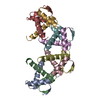
| ||||||||
| Unit cell |
|
-Components
| #1: Protein | Mass: 8972.801 Da / Num. of mol.: 8 Source method: isolated from a genetically manipulated source Source: (gene. exp.) Vibrio cholerae serotype O1 (strain ATCC 39315 / El Tor Inaba N16961) (bacteria)Strain: ATCC 39315 / El Tor Inaba N16961 / Gene: parD, VC_A0360.1 / Production host: |
|---|
-Experimental details
-Experiment
| Experiment | Method: X-RAY DIFFRACTION / Number of used crystals: 1 |
|---|
- Sample preparation
| Crystal | Density Matthews: 2.58 Å3/Da / Density % sol: 52.27 % |
|---|---|
| Crystal grow | Temperature: 293 K / Method: vapor diffusion, sitting drop / pH: 6 Details: 0.2 M Lithium sulfate, 0.1 M MES pH 6 and 20 % w/v PEG 4000 |
-Data collection
| Diffraction | Mean temperature: 100 K / Serial crystal experiment: N |
|---|---|
| Diffraction source | Source: SYNCHROTRON / Site: SOLEIL  / Beamline: PROXIMA 2 / Wavelength: 0.9801 Å / Beamline: PROXIMA 2 / Wavelength: 0.9801 Å |
| Detector | Type: DECTRIS EIGER X 9M / Detector: PIXEL / Date: Sep 6, 2017 |
| Radiation | Protocol: SINGLE WAVELENGTH / Monochromatic (M) / Laue (L): M / Scattering type: x-ray |
| Radiation wavelength | Wavelength: 0.9801 Å / Relative weight: 1 |
| Reflection | Resolution: 3.08→45.95 Å / Num. obs: 8845 / % possible obs: 99.5 % / Redundancy: 6.8 % / CC1/2: 0.996 / Rmerge(I) obs: 0.1738 / Net I/σ(I): 7.36 |
| Reflection shell | Resolution: 3.08→3.19 Å / Rmerge(I) obs: 1.41 / Num. unique obs: 842 / CC1/2: 0.383 |
-Phasing
| Phasing | Method: molecular replacement |
|---|
- Processing
| Software |
| ||||||||||||||||||||||||||||
|---|---|---|---|---|---|---|---|---|---|---|---|---|---|---|---|---|---|---|---|---|---|---|---|---|---|---|---|---|---|
| Refinement | Method to determine structure: MOLECULAR REPLACEMENT Starting model: 3kxe Resolution: 3.08→45.95 Å / SU ML: 0.49 / Cross valid method: THROUGHOUT / σ(F): 1.35 / Phase error: 35.02 / Stereochemistry target values: ML
| ||||||||||||||||||||||||||||
| Solvent computation | Shrinkage radii: 0.9 Å / VDW probe radii: 1.11 Å / Solvent model: FLAT BULK SOLVENT MODEL | ||||||||||||||||||||||||||||
| Displacement parameters | Biso max: 195.24 Å2 / Biso mean: 84.5736 Å2 / Biso min: 52.83 Å2 | ||||||||||||||||||||||||||||
| Refinement step | Cycle: final / Resolution: 3.08→45.95 Å
| ||||||||||||||||||||||||||||
| LS refinement shell | Refine-ID: X-RAY DIFFRACTION / Rfactor Rfree error: 0 / Total num. of bins used: 3
|
