 Movie
Movie Controller

[English] 日本語
 Yorodumi
- PDB-7ycu: Heterotetramer of Antitoxin PrpA together with Toxin PrpT from Ps... -
+ Open data

- Basic information
| Entry | Database: PDB / ID: 7ycu | ||||||
|---|---|---|---|---|---|---|---|
| Title | Heterotetramer of Antitoxin PrpA together with Toxin PrpT from Pseudoalteromonas rubra | ||||||
 Components Components |
| ||||||
 Keywords Keywords | TOXIN/ANTITOXIN / ParE/RelE toxin / ParD antitoxin / RHH / DNA binding motif / Neutralization / ANTITOXIN / TOXIN-ANTITOXIN complex | ||||||
| Function / homology |  Function and homology information Function and homology information | ||||||
| Biological species |  Pseudoalteromonas rubra (bacteria) Pseudoalteromonas rubra (bacteria) | ||||||
| Method |  X-RAY DIFFRACTION / SYNCHROTRON / MOLECULAR REPLACEMENT / Resolution: 1.79 Å X-RAY DIFFRACTION / SYNCHROTRON / MOLECULAR REPLACEMENT / Resolution: 1.79 Å | ||||||
 Authors Authors | Wang, C.C. / Niu, C.Y. / Niu, L.W. | ||||||
| Funding support |  China, 1items China, 1items
| ||||||
 Citation Citation | Journal: Front Microbiol / Year: 2022 Title: Structural insights into the PrpTA toxin-antitoxin system in Pseudoalteromonas rubra. Authors: Wang, C. / Niu, C. / Hidayatullah, K.M. / Xue, L. / Zhu, Z. / Niu, L. | ||||||
| History |
|
- Structure visualization
| Structure viewer | Molecule: MolmilJmol/JSmol |
|---|
- Downloads & links
-Download
| PDBx/mmCIF format | 7ycu.cif.gz | 93.7 KB | Display | PDBx/mmCIF format |
|---|---|---|---|---|
| PDB format | pdb7ycu.ent.gz | 68.8 KB | Display | PDB format |
| PDBx/mmJSON format | 7ycu.json.gz | Tree view | PDBx/mmJSON format | |
| Others |  Other downloads Other downloads |
-Validation report
| Arichive directory | https://data.pdbj.org/pub/pdb/validation_reports/yc/7ycuftp://data.pdbj.org/pub/pdb/validation_reports/yc/7ycu | HTTPS FTP |
|---|
-Related structure data


-Links
 PDBj
PDBj- Assembly
| Deposited unit | 
| ||||||||||||
|---|---|---|---|---|---|---|---|---|---|---|---|---|---|
| 1 | 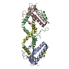
| ||||||||||||
| Unit cell |
|
-Components
| #1: Protein | Mass: 11412.850 Da / Num. of mol.: 2 Source method: isolated from a genetically manipulated source Source: (gene. exp.) Pseudoalteromonas rubra (bacteria) / Gene: AT705_24520 / Production host: #2: Protein | Mass: 11683.994 Da / Num. of mol.: 2 Source method: isolated from a genetically manipulated source Source: (gene. exp.) Pseudoalteromonas rubra (bacteria) / Gene: AT705_24525 / Production host: #3: Water | ChemComp-HOH / |  Mass: 18.015 Da / Num. of mol.: 354 / Source method: isolated from a natural source / Formula: H2O Mass: 18.015 Da / Num. of mol.: 354 / Source method: isolated from a natural source / Formula: H2O |
|---|
-Experimental details
-Experiment
| Experiment | Method: X-RAY DIFFRACTION / Number of used crystals: 1 |
|---|
- Sample preparation
| Crystal | Density Matthews: 2.27 Å3/Da / Density % sol: 45.91 % |
|---|---|
| Crystal grow | Temperature: 289 K / Method: vapor diffusion, sitting drop / pH: 6 / Details: 0.1 M Sodium cacodylate pH 6.0, 15 % w/v PEG 4000 |
-Data collection
| Diffraction | Mean temperature: 100 K / Serial crystal experiment: N |
|---|---|
| Diffraction source | Source: SYNCHROTRON / Site: SSRF / Beamline: BL02U1 / Wavelength: 0.979183 Å |
| Detector | Type: DECTRIS EIGER2 S 9M / Detector: PIXEL / Date: Jan 16, 2022 |
| Radiation | Protocol: SINGLE WAVELENGTH / Monochromatic (M) / Laue (L): M / Scattering type: x-ray |
| Radiation wavelength | Wavelength: 0.979183 Å / Relative weight: 1 |
| Reflection | Resolution: 1.79→44.76 Å / Num. obs: 38264 / % possible obs: 98.64 % / Redundancy: 6.7 % / Biso Wilson estimate: 21.43 Å2 / CC1/2: 0.999 / CC star: 1 / Rmerge(I) obs: 0.08416 / Rpim(I) all: 0.03478 / Rrim(I) all: 0.0912 / Net I/σ(I): 16.7 |
| Reflection shell | Resolution: 1.79→1.857 Å / Redundancy: 5.7 % / Rmerge(I) obs: 0.7912 / Mean I/σ(I) obs: 2.9 / Num. unique obs: 3740 / CC1/2: 0.809 / CC star: 0.946 / Rpim(I) all: 0.3588 / Rrim(I) all: 0.8715 / % possible all: 97.22 |
- Processing
| Software |
| |||||||||||||||||||||||||||||||||||||||||||||||||||||||||||||||||||||||||||||||||||||||||||||||||||||||||
|---|---|---|---|---|---|---|---|---|---|---|---|---|---|---|---|---|---|---|---|---|---|---|---|---|---|---|---|---|---|---|---|---|---|---|---|---|---|---|---|---|---|---|---|---|---|---|---|---|---|---|---|---|---|---|---|---|---|---|---|---|---|---|---|---|---|---|---|---|---|---|---|---|---|---|---|---|---|---|---|---|---|---|---|---|---|---|---|---|---|---|---|---|---|---|---|---|---|---|---|---|---|---|---|---|---|---|
| Refinement | Method to determine structure: MOLECULAR REPLACEMENT / Resolution: 1.79→44.76 Å / SU ML: 0.1801 / Cross valid method: FREE R-VALUE / σ(F): 1.36 / Phase error: 19.2939 Stereochemistry target values: GeoStd + Monomer Library + CDL v1.2
| |||||||||||||||||||||||||||||||||||||||||||||||||||||||||||||||||||||||||||||||||||||||||||||||||||||||||
| Solvent computation | Shrinkage radii: 0.9 Å / VDW probe radii: 1.1 Å / Solvent model: FLAT BULK SOLVENT MODEL | |||||||||||||||||||||||||||||||||||||||||||||||||||||||||||||||||||||||||||||||||||||||||||||||||||||||||
| Displacement parameters | Biso mean: 25.5 Å2 | |||||||||||||||||||||||||||||||||||||||||||||||||||||||||||||||||||||||||||||||||||||||||||||||||||||||||
| Refinement step | Cycle: LAST / Resolution: 1.79→44.76 Å
| |||||||||||||||||||||||||||||||||||||||||||||||||||||||||||||||||||||||||||||||||||||||||||||||||||||||||
| Refine LS restraints |
| |||||||||||||||||||||||||||||||||||||||||||||||||||||||||||||||||||||||||||||||||||||||||||||||||||||||||
| LS refinement shell |
|
