 Movie
Movie Controller

[English] 日本語
 Yorodumi
- PDB-7y9t: Structure of the auxin exporter PIN1 in Arabidopsis thaliana in t... -
+ Open data

- Basic information
| Entry | Database: PDB / ID: 7y9t | |||||||||||||||
|---|---|---|---|---|---|---|---|---|---|---|---|---|---|---|---|---|
| Title | Structure of the auxin exporter PIN1 in Arabidopsis thaliana in the apo state | |||||||||||||||
 Components Components |
| |||||||||||||||
 Keywords Keywords | TRANSPORT PROTEIN | |||||||||||||||
| Function / homology |  Function and homology information Function and homology informationER body / cotyledon morphogenesis / leaf formation / cotyledon development / leaf shaping / adventitious root development / shoot system development / xylem and phloem pattern formation / somatic embryogenesis / auxin transport ...ER body / cotyledon morphogenesis / leaf formation / cotyledon development / leaf shaping / adventitious root development / shoot system development / xylem and phloem pattern formation / somatic embryogenesis / auxin transport / gravitropism / auxin export across the plasma membrane / auxin efflux transmembrane transporter activity / inflorescence development / flower development / auxin polar transport / root development / photomorphogenesis / plant-type cell wall / embryo development ending in seed dormancy / auxin-activated signaling pathway / plasmodesma / basal plasma membrane / cell periphery / apical part of cell / apical plasma membrane / endoplasmic reticulum / protein homodimerization activity / identical protein binding / plasma membrane / cytoplasm Similarity search - Function | |||||||||||||||
| Biological species |   | |||||||||||||||
| Method | ELECTRON MICROSCOPY / single particle reconstruction / cryo EM / Resolution: 3.1 Å | |||||||||||||||
 Authors Authors | Sun, L. / Liu, X. / Yang, Z. / Xia, J. | |||||||||||||||
| Funding support |  China, 4items China, 4items
| |||||||||||||||
 Citation Citation | Journal: Nature / Year: 2022 Title: Structural insights into auxin recognition and efflux by Arabidopsis PIN1. Authors: Zhisen Yang / Jing Xia / Jingjing Hong / Chenxi Zhang / Hong Wei / Wei Ying / Chunqiao Sun / Lianghanxiao Sun / Yanbo Mao / Yongxiang Gao / Shutang Tan / Jiří Friml / Dianfan Li / Xin Liu / Linfeng Sun /  Abstract: Polar auxin transport is unique to plants and coordinates their growth and development. The PIN-FORMED (PIN) auxin transporters exhibit highly asymmetrical localizations at the plasma membrane and ...Polar auxin transport is unique to plants and coordinates their growth and development. The PIN-FORMED (PIN) auxin transporters exhibit highly asymmetrical localizations at the plasma membrane and drive polar auxin transport; however, their structures and transport mechanisms remain largely unknown. Here, we report three inward-facing conformation structures of Arabidopsis thaliana PIN1: the apo state, bound to the natural auxin indole-3-acetic acid (IAA), and in complex with the polar auxin transport inhibitor N-1-naphthylphthalamic acid (NPA). The transmembrane domain of PIN1 shares a conserved NhaA fold. In the substrate-bound structure, IAA is coordinated by both hydrophobic stacking and hydrogen bonding. NPA competes with IAA for the same site at the intracellular pocket, but with a much higher affinity. These findings inform our understanding of the substrate recognition and transport mechanisms of PINs and set up a framework for future research on directional auxin transport, one of the most crucial processes underlying plant development. | |||||||||||||||
| History |
|
- Structure visualization
| Structure viewer | Molecule: MolmilJmol/JSmol |
|---|
- Downloads & links
-Download
| PDBx/mmCIF format | 7y9t.cif.gz | 186.3 KB | Display | PDBx/mmCIF format |
|---|---|---|---|---|
| PDB format | pdb7y9t.ent.gz | 143.9 KB | Display | PDB format |
| PDBx/mmJSON format | 7y9t.json.gz | Tree view | PDBx/mmJSON format | |
| Others |  Other downloads Other downloads |
-Validation report
| Arichive directory | https://data.pdbj.org/pub/pdb/validation_reports/y9/7y9tftp://data.pdbj.org/pub/pdb/validation_reports/y9/7y9t | HTTPS FTP |
|---|
-Related structure data
| Related structure data |  33691MC  7y9uC 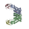 7y9vC M: map data used to model this data C: citing same article ( |
|---|---|
| Similar structure data |
-Links
 PDBj
PDBj

- Assembly
| Deposited unit | 
|
|---|---|
| 1 |
|
-Components
| #1: Protein | Mass: 67080.781 Da / Num. of mol.: 2 Source method: isolated from a genetically manipulated source Source: (gene. exp.)  Homo sapiens (human) / References: UniProt: Q9C6B8 Homo sapiens (human) / References: UniProt: Q9C6B8#2: Antibody | Mass: 13369.910 Da / Num. of mol.: 2 Source method: isolated from a genetically manipulated source Source: (gene. exp.) Has protein modification | Y | |
|---|
-Experimental details
-Experiment
| Experiment | Method: ELECTRON MICROSCOPY |
|---|---|
| EM experiment | Aggregation state: PARTICLE / 3D reconstruction method: single particle reconstruction |
- Sample preparation
| Component |
| ||||||||||||||||||||||||
|---|---|---|---|---|---|---|---|---|---|---|---|---|---|---|---|---|---|---|---|---|---|---|---|---|---|
| Molecular weight | Experimental value: NO | ||||||||||||||||||||||||
| Source (natural) |
| ||||||||||||||||||||||||
| Source (recombinant) |
| ||||||||||||||||||||||||
| Buffer solution | pH: 7.4 | ||||||||||||||||||||||||
| Specimen | Embedding applied: NO / Shadowing applied: NO / Staining applied: NO / Vitrification applied: YES | ||||||||||||||||||||||||
| Vitrification | Cryogen name: ETHANE |
- Electron microscopy imaging
| Experimental equipment |  Model: Titan Krios / Image courtesy: FEI Company |
|---|---|
| Microscopy | Model: FEI TITAN KRIOS |
| Electron gun | Electron source:  FIELD EMISSION GUN / Accelerating voltage: 300 kV / Illumination mode: SPOT SCAN FIELD EMISSION GUN / Accelerating voltage: 300 kV / Illumination mode: SPOT SCAN |
| Electron lens | Mode: DIFFRACTION / Nominal defocus max: 2300 nm / Nominal defocus min: 1500 nm |
| Image recording | Electron dose: 50 e/Å2 / Film or detector model: GATAN K3 BIOQUANTUM (6k x 4k) |
- Processing
| CTF correction | Type: NONE |
|---|---|
| 3D reconstruction | Resolution: 3.1 Å / Resolution method: FSC 0.143 CUT-OFF / Num. of particles: 210597 / Symmetry type: POINT |
