 Movie
Movie Controller

[English] 日本語
 Yorodumi
- PDB-7y8l: Structure of ScIRED-R2-V3 from Streptomyces clavuligerus in compl... -
+ Open data

- Basic information
| Entry | Database: PDB / ID: 7y8l | ||||||
|---|---|---|---|---|---|---|---|
| Title | Structure of ScIRED-R2-V3 from Streptomyces clavuligerus in complex with 5-(2,5-difluorophenyl)-3,4-dihydro-2H-pyrrole | ||||||
 Components Components | reductase for protein | ||||||
 Keywords Keywords | OXIDOREDUCTASE/INHIBITOR / substrate binding / OXIDOREDUCTASE-inhibitor complex | ||||||
| Function / homology | Chem-4IS / NADP NICOTINAMIDE-ADENINE-DINUCLEOTIDE PHOSPHATE / Chem-NDP Function and homology information Function and homology information | ||||||
| Biological species |  Streptomyces clavuligerus (bacteria) Streptomyces clavuligerus (bacteria) | ||||||
| Method |  X-RAY DIFFRACTION / MOLECULAR REPLACEMENT / Resolution: 2.41 Å X-RAY DIFFRACTION / MOLECULAR REPLACEMENT / Resolution: 2.41 Å | ||||||
 Authors Authors | Zhang, L.L. / Liu, W.D. / Shi, M. / Huang, J.W. / Yang, Y. / Chen, C.C. / Guo, R.T. | ||||||
| Funding support | 1items
| ||||||
 Citation Citation | Journal: Acs Catalysis / Year: 2022 Title: Engineered Imine Reductase for Larotrectinib Intermediate Manufacture Authors: Chen, Q. / Li, B.B. / Zhang, L. / Chen, X.R. / Zhu, X.X. / Chen, F.F. / Shi, M. / Chen, C.C. / Yang, Y. / Guo, R.T. / Liu, W. / Xu, J.H. / Zheng, G.W. | ||||||
| History |
|
- Structure visualization
| Structure viewer | Molecule: MolmilJmol/JSmol |
|---|
- Downloads & links
-Download
| PDBx/mmCIF format | 7y8l.cif.gz | 224.5 KB | Display | PDBx/mmCIF format |
|---|---|---|---|---|
| PDB format | pdb7y8l.ent.gz | 181.2 KB | Display | PDB format |
| PDBx/mmJSON format | 7y8l.json.gz | Tree view | PDBx/mmJSON format | |
| Others |  Other downloads Other downloads |
-Validation report
| Arichive directory | https://data.pdbj.org/pub/pdb/validation_reports/y8/7y8lftp://data.pdbj.org/pub/pdb/validation_reports/y8/7y8l | HTTPS FTP |
|---|
-Related structure data
| Related structure data |  7y8kC 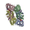 7y8mC 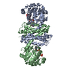 7y8nC  4oqyS S: Starting model for refinement C: citing same article ( |
|---|---|
| Similar structure data |
-Links
 PDBj
PDBj

- Assembly
| Deposited unit | 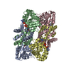
| |||||||||||||||||||||||||||||||||||||||||||||||||||||||||||||||||
|---|---|---|---|---|---|---|---|---|---|---|---|---|---|---|---|---|---|---|---|---|---|---|---|---|---|---|---|---|---|---|---|---|---|---|---|---|---|---|---|---|---|---|---|---|---|---|---|---|---|---|---|---|---|---|---|---|---|---|---|---|---|---|---|---|---|---|
| 1 |
| |||||||||||||||||||||||||||||||||||||||||||||||||||||||||||||||||
| 2 | 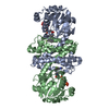
| |||||||||||||||||||||||||||||||||||||||||||||||||||||||||||||||||
| 3 | 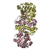
| |||||||||||||||||||||||||||||||||||||||||||||||||||||||||||||||||
| Unit cell |
| |||||||||||||||||||||||||||||||||||||||||||||||||||||||||||||||||
| Noncrystallographic symmetry (NCS) | NCS domain:
NCS domain segments: Component-ID: 1 / Ens-ID: 1
|
-Components
| #1: Protein | Mass: 30523.529 Da / Num. of mol.: 4 Source method: isolated from a genetically manipulated source Source: (gene. exp.) Streptomyces clavuligerus (bacteria) / Plasmid: pET28 / Production host: #2: Chemical |   Mass: 745.421 Da / Num. of mol.: 3 / Source method: obtained synthetically / Formula: C21H30N7O17P3 / Feature type: SUBJECT OF INVESTIGATION Mass: 745.421 Da / Num. of mol.: 3 / Source method: obtained synthetically / Formula: C21H30N7O17P3 / Feature type: SUBJECT OF INVESTIGATION#3: Chemical | ChemComp-4IS /   Mass: 181.182 Da / Num. of mol.: 4 / Source method: obtained synthetically / Formula: C10H9F2N / Feature type: SUBJECT OF INVESTIGATION Mass: 181.182 Da / Num. of mol.: 4 / Source method: obtained synthetically / Formula: C10H9F2N / Feature type: SUBJECT OF INVESTIGATION#4: Chemical | ChemComp-NAP / |   Mass: 743.405 Da / Num. of mol.: 1 / Source method: obtained synthetically / Formula: C21H28N7O17P3 Mass: 743.405 Da / Num. of mol.: 1 / Source method: obtained synthetically / Formula: C21H28N7O17P3#5: Water | ChemComp-HOH / |  Mass: 18.015 Da / Num. of mol.: 128 / Source method: isolated from a natural source / Formula: H2O Mass: 18.015 Da / Num. of mol.: 128 / Source method: isolated from a natural source / Formula: H2OHas ligand of interest | Y | |
|---|
-Experimental details
-Experiment
| Experiment | Method: X-RAY DIFFRACTION / Number of used crystals: 1 |
|---|
- Sample preparation
| Crystal | Density Matthews: 2.19 Å3/Da / Density % sol: 43.94 % / Mosaicity: 0 ° |
|---|---|
| Crystal grow | Temperature: 298 K / Method: vapor diffusion, sitting drop / pH: 6.5 Details: 12% Polyethylene glycol 8000, 0.1 M 2-(4-Morpholino) ethanesulfonic acid pH 6.0, 0.1M Calcium Acetate |
-Data collection
| Diffraction | Mean temperature: 100 K / Serial crystal experiment: N |
|---|---|
| Diffraction source | Source: LIQUID ANODE / Type: BRUKER METALJET / Wavelength: 1.34138 Å |
| Detector | Type: Bruker PHOTON III / Detector: PIXEL / Date: Feb 7, 2021 |
| Radiation | Protocol: SINGLE WAVELENGTH / Monochromatic (M) / Laue (L): M / Scattering type: x-ray |
| Radiation wavelength | Wavelength: 1.34138 Å / Relative weight: 1 |
| Reflection | Resolution: 2.41→36.42 Å / Num. obs: 40850 / % possible obs: 99.1 % / Redundancy: 6.87 % / Rmerge(I) obs: 0.118 / Net I/σ(I): 8.23 |
| Reflection shell | Resolution: 2.41→2.51 Å / Redundancy: 4.9 % / Rmerge(I) obs: 0.47 / Num. unique obs: 4542 / % possible all: 96.8 |
- Processing
| Software |
| |||||||||||||||||||||||||||||||||||||||||||||||||||||||||||||||||||||||||||||
|---|---|---|---|---|---|---|---|---|---|---|---|---|---|---|---|---|---|---|---|---|---|---|---|---|---|---|---|---|---|---|---|---|---|---|---|---|---|---|---|---|---|---|---|---|---|---|---|---|---|---|---|---|---|---|---|---|---|---|---|---|---|---|---|---|---|---|---|---|---|---|---|---|---|---|---|---|---|---|
| Refinement | Method to determine structure: MOLECULAR REPLACEMENT Starting model: 4OQY Resolution: 2.41→36.42 Å / SU ML: 0.33 / Cross valid method: THROUGHOUT / σ(F): 1.34 / Phase error: 30.12 / Stereochemistry target values: ML
| |||||||||||||||||||||||||||||||||||||||||||||||||||||||||||||||||||||||||||||
| Solvent computation | Shrinkage radii: 0.9 Å / VDW probe radii: 1.11 Å / Solvent model: FLAT BULK SOLVENT MODEL | |||||||||||||||||||||||||||||||||||||||||||||||||||||||||||||||||||||||||||||
| Displacement parameters | Biso max: 129.29 Å2 / Biso mean: 43.47 Å2 / Biso min: 16.61 Å2 | |||||||||||||||||||||||||||||||||||||||||||||||||||||||||||||||||||||||||||||
| Refinement step | Cycle: final / Resolution: 2.41→36.42 Å
| |||||||||||||||||||||||||||||||||||||||||||||||||||||||||||||||||||||||||||||
| Refine LS restraints NCS |
| |||||||||||||||||||||||||||||||||||||||||||||||||||||||||||||||||||||||||||||
| LS refinement shell | Refine-ID: X-RAY DIFFRACTION / Rfactor Rfree error: 0 / Total num. of bins used: 10
|
