 Movie
Movie Controller


+ Open data

- Basic information
| Entry | Database: PDB / ID: 7y4q | ||||||||||||
|---|---|---|---|---|---|---|---|---|---|---|---|---|---|
| Title | Semaphorin 6D in complex with Plexin A1 | ||||||||||||
 Components Components |
| ||||||||||||
 Keywords Keywords | SIGNALING PROTEIN / signaling complex | ||||||||||||
| Function / homology |  Function and homology information Function and homology informationOther semaphorin interactions / olfactory nerve formation / neuron projection guidance / dichotomous subdivision of terminal units involved in salivary gland branching / gonadotrophin-releasing hormone neuronal migration to the hypothalamus / semaphorin receptor binding / Other semaphorin interactions / T cell activation via T cell receptor contact with antigen bound to MHC molecule on antigen presenting cell / negative regulation of smooth muscle cell migration / semaphorin receptor complex ...Other semaphorin interactions / olfactory nerve formation / neuron projection guidance / dichotomous subdivision of terminal units involved in salivary gland branching / gonadotrophin-releasing hormone neuronal migration to the hypothalamus / semaphorin receptor binding / Other semaphorin interactions / T cell activation via T cell receptor contact with antigen bound to MHC molecule on antigen presenting cell / negative regulation of smooth muscle cell migration / semaphorin receptor complex / ventricular system development / SEMA3A-Plexin repulsion signaling by inhibiting Integrin adhesion / semaphorin receptor activity / chemorepellent activity / CRMPs in Sema3A signaling / regulation of smooth muscle cell migration / RHOD GTPase cycle / RND1 GTPase cycle / neural crest cell migration / neuron projection extension / positive regulation of smooth muscle cell migration / smooth muscle cell migration / semaphorin-plexin signaling pathway / Sema3A PAK dependent Axon repulsion / synapse assembly / axon guidance / regulation of cell migration / positive regulation of cell migration / receptor ligand activity / glutamatergic synapse / Golgi apparatus / extracellular exosome / nucleoplasm / plasma membrane / cytosol / cytoplasm Similarity search - Function | ||||||||||||
| Biological species |  Homo sapiens (human) Homo sapiens (human) | ||||||||||||
| Method |  X-RAY DIFFRACTION / SYNCHROTRON / MOLECULAR REPLACEMENT / Resolution: 4.7 Å X-RAY DIFFRACTION / SYNCHROTRON / MOLECULAR REPLACEMENT / Resolution: 4.7 Å | ||||||||||||
 Authors Authors | Tanaka, T. / Neyazaki, M. / Nogi, T. | ||||||||||||
| Funding support |  Japan, 3items Japan, 3items
| ||||||||||||
 Citation Citation | Journal: Protein Sci. / Year: 2022 Title: Hybrid in vitro/in silico analysis of low-affinity protein-protein interactions that regulate signal transduction by Sema6D. Authors: Tanaka, T. / Ekimoto, T. / Nagatomo, M. / Neyazaki, M. / Shimoji, E. / Yamane, T. / Kanagawa, S. / Oi, R. / Mihara, E. / Takagi, J. / Akashi, S. / Ikeguchi, M. / Nogi, T. | ||||||||||||
| History |
|
- Structure visualization
| Structure viewer | Molecule: MolmilJmol/JSmol |
|---|
- Downloads & links
-Download
| PDBx/mmCIF format | 7y4q.cif.gz | 595.3 KB | Display | PDBx/mmCIF format |
|---|---|---|---|---|
| PDB format | pdb7y4q.ent.gz | 390.3 KB | Display | PDB format |
| PDBx/mmJSON format | 7y4q.json.gz | Tree view | PDBx/mmJSON format | |
| Others |  Other downloads Other downloads |
-Validation report
| Arichive directory | https://data.pdbj.org/pub/pdb/validation_reports/y4/7y4qftp://data.pdbj.org/pub/pdb/validation_reports/y4/7y4q | HTTPS FTP |
|---|
-Related structure data
| Related structure data |  7y4oC 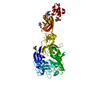 7y4pC  7cyt 7d07 S: Starting model for refinement C: citing same article ( |
|---|---|
| Similar structure data |
-Links
 PDBj
PDBj


- Assembly
| Deposited unit | 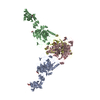
| ||||||||||||
|---|---|---|---|---|---|---|---|---|---|---|---|---|---|
| 1 |
| ||||||||||||
| Unit cell |
|
-Components
| #1: Protein | Mass: 77079.102 Da / Num. of mol.: 2 / Fragment: ectodomain fragment Source method: isolated from a genetically manipulated source Source: (gene. exp.) Homo sapiens (human) / Gene: PLXNA1, NOV, PLXN1 / Cell line (production host): HEK293S GnT1- / Production host: Homo sapiens (human) / References: UniProt: Q9UIW2#2: Protein | Mass: 62522.633 Da / Num. of mol.: 2 / Fragment: ectodomain fragment / Mutation: S332G Source method: isolated from a genetically manipulated source Source: (gene. exp.)  Cricetulus griseus (Chinese hamster) / References: UniProt: A0A0G2JZC4 Cricetulus griseus (Chinese hamster) / References: UniProt: A0A0G2JZC4#3: Polysaccharide | 2-acetamido-2-deoxy-beta-D-glucopyranose-(1-4)-2-acetamido-2-deoxy-beta-D-glucopyranose Source method: isolated from a genetically manipulated source #4: Sugar | ChemComp-NAG /   Type: D-saccharide, beta linking / Mass: 221.208 Da / Num. of mol.: 4 / Source method: obtained synthetically / Formula: C8H15NO6 / Feature type: SUBJECT OF INVESTIGATION Type: D-saccharide, beta linking / Mass: 221.208 Da / Num. of mol.: 4 / Source method: obtained synthetically / Formula: C8H15NO6 / Feature type: SUBJECT OF INVESTIGATIONHas ligand of interest | Y | Has protein modification | Y | |
|---|
-Experimental details
-Experiment
| Experiment | Method: X-RAY DIFFRACTION / Number of used crystals: 1 |
|---|
- Sample preparation
| Crystal | Density Matthews: 9.82 Å3/Da / Density % sol: 87.48 % |
|---|---|
| Crystal grow | Temperature: 283 K / Method: vapor diffusion, hanging drop / pH: 7.4 / Details: 150 mM NaCl and 10 mM Tris-Cl (pH 7.4) |
-Data collection
| Diffraction | Mean temperature: 100 K / Serial crystal experiment: N |
|---|---|
| Diffraction source | Source: SYNCHROTRON / Site: Photon Factory / Beamline: BL-17A / Wavelength: 0.98 Å |
| Detector | Type: DECTRIS PILATUS3 S 6M / Detector: PIXEL / Date: Jun 14, 2016 |
| Radiation | Protocol: SINGLE WAVELENGTH / Monochromatic (M) / Laue (L): M / Scattering type: x-ray |
| Radiation wavelength | Wavelength: 0.98 Å / Relative weight: 1 |
| Reflection | Resolution: 4.7→49.43 Å / Num. obs: 110174 / % possible obs: 99.2 % / Redundancy: 3.4 % / Biso Wilson estimate: 155.92 Å2 / Rmerge(I) obs: 0.17 / Net I/σ(I): 6.1 |
| Reflection shell | Resolution: 4.7→4.84 Å / Redundancy: 3.5 % / Rmerge(I) obs: 0.938 / Mean I/σ(I) obs: 1.5 / Num. unique obs: 4580 / % possible all: 99.8 |
- Processing
| Software |
| |||||||||||||||||||||||||||||||||||||||||||||||||||||||||||||||||||||||||||||||||||||||||||||||||||||||||||||||||||||||||||||||||||||||||||||||||||||||||||||||||||||||||||||||||||||||||||||||||||||||||||||||||||||||||
|---|---|---|---|---|---|---|---|---|---|---|---|---|---|---|---|---|---|---|---|---|---|---|---|---|---|---|---|---|---|---|---|---|---|---|---|---|---|---|---|---|---|---|---|---|---|---|---|---|---|---|---|---|---|---|---|---|---|---|---|---|---|---|---|---|---|---|---|---|---|---|---|---|---|---|---|---|---|---|---|---|---|---|---|---|---|---|---|---|---|---|---|---|---|---|---|---|---|---|---|---|---|---|---|---|---|---|---|---|---|---|---|---|---|---|---|---|---|---|---|---|---|---|---|---|---|---|---|---|---|---|---|---|---|---|---|---|---|---|---|---|---|---|---|---|---|---|---|---|---|---|---|---|---|---|---|---|---|---|---|---|---|---|---|---|---|---|---|---|---|---|---|---|---|---|---|---|---|---|---|---|---|---|---|---|---|---|---|---|---|---|---|---|---|---|---|---|---|---|---|---|---|---|---|---|---|---|---|---|---|---|---|---|---|---|---|---|---|---|
| Refinement | Method to determine structure: MOLECULAR REPLACEMENT Starting model: 7D07, 7CYT Resolution: 4.7→49.43 Å / SU ML: 0.6709 / Cross valid method: FREE R-VALUE / σ(F): 0.05 / Phase error: 27.178 / Stereochemistry target values: GeoStd + Monomer Library
| |||||||||||||||||||||||||||||||||||||||||||||||||||||||||||||||||||||||||||||||||||||||||||||||||||||||||||||||||||||||||||||||||||||||||||||||||||||||||||||||||||||||||||||||||||||||||||||||||||||||||||||||||||||||||
| Solvent computation | Shrinkage radii: 0.9 Å / VDW probe radii: 1.11 Å / Solvent model: FLAT BULK SOLVENT MODEL | |||||||||||||||||||||||||||||||||||||||||||||||||||||||||||||||||||||||||||||||||||||||||||||||||||||||||||||||||||||||||||||||||||||||||||||||||||||||||||||||||||||||||||||||||||||||||||||||||||||||||||||||||||||||||
| Displacement parameters | Biso mean: 200.63 Å2 | |||||||||||||||||||||||||||||||||||||||||||||||||||||||||||||||||||||||||||||||||||||||||||||||||||||||||||||||||||||||||||||||||||||||||||||||||||||||||||||||||||||||||||||||||||||||||||||||||||||||||||||||||||||||||
| Refinement step | Cycle: LAST / Resolution: 4.7→49.43 Å
| |||||||||||||||||||||||||||||||||||||||||||||||||||||||||||||||||||||||||||||||||||||||||||||||||||||||||||||||||||||||||||||||||||||||||||||||||||||||||||||||||||||||||||||||||||||||||||||||||||||||||||||||||||||||||
| Refine LS restraints |
| |||||||||||||||||||||||||||||||||||||||||||||||||||||||||||||||||||||||||||||||||||||||||||||||||||||||||||||||||||||||||||||||||||||||||||||||||||||||||||||||||||||||||||||||||||||||||||||||||||||||||||||||||||||||||
| LS refinement shell |
|
