 Movie
Movie Controller

[English] 日本語
 Yorodumi
- PDB-7y1b: 3.2 angstrom cryo-EM structure of extracellular region of mouse B... -
+ Open data

- Basic information
| Entry | Database: PDB / ID: 7y1b | |||||||||
|---|---|---|---|---|---|---|---|---|---|---|
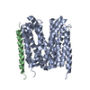
| Title | 3.2 angstrom cryo-EM structure of extracellular region of mouse Basigin-2 in complex with the Fab fragment of antibody 6E7F1 | |||||||||
 Components Components |
| |||||||||
 Keywords Keywords | TRANSPORT PROTEIN / Transporter / glucose metabolism / monocarboxylic acid transport / alpha helical TMs | |||||||||
| Function / homology |  Function and homology information Function and homology informationProton-coupled monocarboxylate transport / Aspirin ADME / Basigin interactions / positive regulation of matrix metallopeptidase secretion / acrosomal membrane / Degradation of the extracellular matrix / dendrite self-avoidance / Integrin cell surface interactions / endothelial tube morphogenesis / cell-cell adhesion mediator activity ...Proton-coupled monocarboxylate transport / Aspirin ADME / Basigin interactions / positive regulation of matrix metallopeptidase secretion / acrosomal membrane / Degradation of the extracellular matrix / dendrite self-avoidance / Integrin cell surface interactions / endothelial tube morphogenesis / cell-cell adhesion mediator activity / response to mercury ion / neural retina development / photoreceptor cell maintenance / neuron projection extension / odontogenesis of dentin-containing tooth / D-mannose binding / homophilic cell-cell adhesion / decidualization / positive regulation of vascular endothelial growth factor production / photoreceptor outer segment / response to cAMP / neutrophil chemotaxis / photoreceptor inner segment / positive regulation of endothelial cell migration / embryo implantation / axon guidance / protein localization to plasma membrane / sarcolemma / response to peptide hormone / positive regulation of interleukin-6 production / signaling receptor activity / virus receptor activity / angiogenesis / spermatogenesis / basolateral plasma membrane / positive regulation of viral entry into host cell / endosome / axon / endoplasmic reticulum membrane / plasma membrane Similarity search - Function | |||||||||
| Biological species |  | |||||||||
| Method | ELECTRON MICROSCOPY / single particle reconstruction / cryo EM / Resolution: 3.23 Å | |||||||||
 Authors Authors | Zhang, H. / Yang, X. / Xue, Y. / Huang, Y. / Mo, X. / Zhang, H. / Li, N. / Gao, N. / Li, X. / Wang, S. ...Zhang, H. / Yang, X. / Xue, Y. / Huang, Y. / Mo, X. / Zhang, H. / Li, N. / Gao, N. / Li, X. / Wang, S. / Gao, Y. / Liao, J. | |||||||||
| Funding support |  China, 2items China, 2items
| |||||||||
 Citation Citation | Journal: To Be Published Title: Allosteric modulation of monocarboxylate transporters 1 and 4 by targeting their chaperon Basigin Authors: Zhang, H. / Yang, X. / Xue, Y. / Huang, Y. / Mo, Y. / Zhang, H. / Li, N. / Gao, N. / Li, X. / Wang, S. / Gao, Y. / Liao, J. | |||||||||
| History |
|
- Structure visualization
| Structure viewer | Molecule: MolmilJmol/JSmol |
|---|
- Downloads & links
-Download
| PDBx/mmCIF format | 7y1b.cif.gz | 107.2 KB | Display | PDBx/mmCIF format |
|---|---|---|---|---|
| PDB format | pdb7y1b.ent.gz | 79.2 KB | Display | PDB format |
| PDBx/mmJSON format | 7y1b.json.gz | Tree view | PDBx/mmJSON format | |
| Others |  Other downloads Other downloads |
-Validation report
| Arichive directory | https://data.pdbj.org/pub/pdb/validation_reports/y1/7y1bftp://data.pdbj.org/pub/pdb/validation_reports/y1/7y1b | HTTPS FTP |
|---|
-Related structure data
| Related structure data |  33559MC M: map data used to model this data C: citing same article ( |
|---|---|
| Similar structure data |
-Links
 PDBj
PDBj


- Assembly
| Deposited unit | 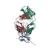
|
|---|---|
| 1 |
|
-Components
| #1: Protein | Mass: 29707.447 Da / Num. of mol.: 1 Source method: isolated from a genetically manipulated source Source: (gene. exp.)  Homo sapiens (human) / References: UniProt: P18572 Homo sapiens (human) / References: UniProt: P18572 |
|---|---|
| #2: Antibody | Mass: 25275.400 Da / Num. of mol.: 1 Source method: isolated from a genetically manipulated source Source: (gene. exp.) |
| #3: Antibody | Mass: 25924.785 Da / Num. of mol.: 1 Source method: isolated from a genetically manipulated source Source: (gene. exp.) |
| Has protein modification | Y |
-Experimental details
-Experiment
| Experiment | Method: ELECTRON MICROSCOPY |
|---|---|
| EM experiment | Aggregation state: PARTICLE / 3D reconstruction method: single particle reconstruction |
- Sample preparation
| Component | Name: 3.2 angstrom cryo-EM structure of extracellular region of mouse MCT1/Basigin-2 in complex with its monoclonal antibody 6E7F1 Type: COMPLEX / Entity ID: all / Source: RECOMBINANT | |||||||||||||||
|---|---|---|---|---|---|---|---|---|---|---|---|---|---|---|---|---|
| Molecular weight | Value: 0.1 MDa / Experimental value: YES | |||||||||||||||
| Source (natural) | Organism: | |||||||||||||||
| Source (recombinant) | Organism: Homo sapiens (human) / Cell: HEK293S / Plasmid: pEG BacMam | |||||||||||||||
| Buffer solution | pH: 7.5 / Details: 20 mM HEPES pH7.5, 150 mM NaCl | |||||||||||||||
| Buffer component |
| |||||||||||||||
| Specimen | Conc.: 0.7 mg/ml / Embedding applied: NO / Shadowing applied: NO / Staining applied: NO / Vitrification applied: YES / Details: This sample is monodisperse | |||||||||||||||
| Specimen support | Grid material: GOLD / Grid mesh size: 300 divisions/in. / Grid type: Quantifoil R1.2/1.3 | |||||||||||||||
| Vitrification | Instrument: FEI VITROBOT MARK IV / Cryogen name: ETHANE / Humidity: 100 % / Chamber temperature: 283 K / Details: blot for 3.5-4.5 seconds before plunging |
- Electron microscopy imaging
| Experimental equipment |  Model: Titan Krios / Image courtesy: FEI Company |
|---|---|
| Microscopy | Model: FEI TITAN KRIOS |
| Electron gun | Electron source:  FIELD EMISSION GUN / Accelerating voltage: 300 kV / Illumination mode: SPOT SCAN FIELD EMISSION GUN / Accelerating voltage: 300 kV / Illumination mode: SPOT SCAN |
| Electron lens | Mode: DIFFRACTION / Nominal defocus max: 2000 nm / Nominal defocus min: 1000 nm / Cs: 2.7 mm |
| Specimen holder | Cryogen: NITROGEN |
| Image recording | Average exposure time: 2.5 sec. / Electron dose: 75 e/Å2 / Detector mode: SUPER-RESOLUTION / Film or detector model: GATAN K2 SUMMIT (4k x 4k) / Num. of real images: 2842 |
| Image scans | Sampling size: 1.9 µm / Width: 1034 / Height: 1034 / Movie frames/image: 48 |
- Processing
| EM software |
| ||||||||||||||||||||||||||||||||||||
|---|---|---|---|---|---|---|---|---|---|---|---|---|---|---|---|---|---|---|---|---|---|---|---|---|---|---|---|---|---|---|---|---|---|---|---|---|---|
| CTF correction | Type: PHASE FLIPPING AND AMPLITUDE CORRECTION | ||||||||||||||||||||||||||||||||||||
| Particle selection | Num. of particles selected: 712923 | ||||||||||||||||||||||||||||||||||||
| 3D reconstruction | Resolution: 3.23 Å / Resolution method: FSC 0.143 CUT-OFF / Num. of particles: 106160 / Symmetry type: POINT | ||||||||||||||||||||||||||||||||||||
| Atomic model building | B value: 39.46 / Protocol: AB INITIO MODEL / Space: REAL / Target criteria: Correlation coefficient |
